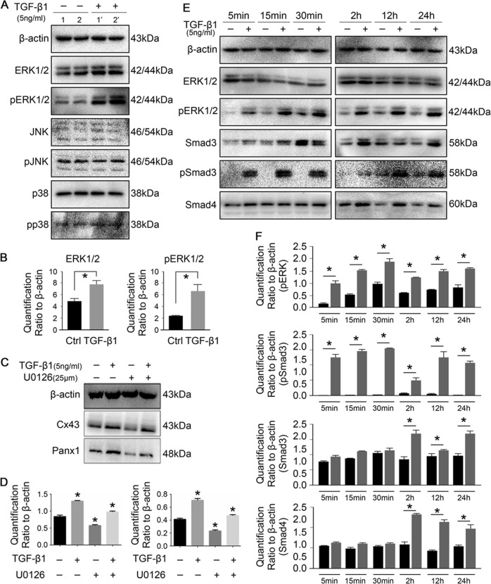
Fig. 4. TGF-β1 mediates gap junctions through ERK and Smad3/4 signaling pathways.
a Representative Western blots showing the different expressions of MAPKs in osteocytes (MLO-Y4 cell line) induced by TGF-β1. Sample 1, 1′ and 2, 2′ are from two independent experiments. b Quantitative analysis showing TGF-β1 induced highly expressions of total and phosphorylated ERK1/2 in osteocytes. Data are presented as the mean ± s.d.; n = 3, *p < 0.05 by t-test. c Representative Western blot images of Cx43 and panx1 showing interaction between TGF-β1 and ERK1/2 signaling. The presence of U0126, ERK1/2 inhibitor, reduced the expression of Cx43 and panx1 at 25 μM. While TGF-β1 promoted the expression of Cx43 and panx1 in the presence of U0126. The results shown are based on three independent experiments (n = 3). d Quantification in c was performed to confirm the protein changes. The results were normalized to β-actin as Western blot loading control. Data are presented as the mean ± s.d.; n = 3, *p < 0.05 by t-test. e Representative Western blots showing protein expressions of total and phosphorylated Smad3 and Smad4 beyond ERK1/2 in osteocytes after treatment with TGF-β1 (5 ng/ml) at different time points. The results were based on three independent experiments (n = 3). f Quantitative analysis of proteins in e. The results were normalized to β-actin as Western blot loading control. Data are presented as the mean ± s.d.; n = 3, *p < 0.05 by t-test

