
Fig. 1. iNKT cells promote CD8+ T cell activation in the absence of exogenous NKT cell antigens.
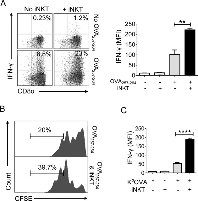
a Sorted OT-1 CD8+ T cells were activated by CD1d knockout DCs (CD1dKO DCs) pretreated with OVA257–264 peptides in the presence or absence of iNKT cells. After two days of stimulation, the production of IFN-γ by CTLs was examined via intracellular FACS analysis. Detection of IFN-γ-producing CD8+ T cells by staining with anti-TCRVβ5.1, anti-CD8α, and anti-IFN-γ (left panel), b mean fluorescence intensity (MFI) of IFN-γ in OT-1 CD8+ T cells (right panel). c CFSE-labeled OT-1 CD8+ T cells were stimulated as described in (a). Representative histogram displaying CFSE dilution assay of OT-1 cells. d OT-1 CD8+ T cells were stimulated with plates coated with KbOVA in the presence or absence of iNKT cells. After 2 days of stimulation, the amount of IFN-γ produced by OT-1 cells was determined. All data shown are representative of four independent experiments with similar results. Data are presented as the mean ± SEM; **p < 0.01 and ****p < 0.0001. One-way ANOVA with Bonferroni’s multiple comparison test was used for data analysis