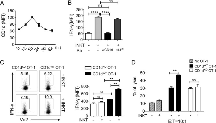
Fig. 2. iNKT cells promote the effector function of CTLs via direct interaction with CD1d on CTLs.
a OT-1 CD8+ T cells were stimulated with OVA257–264-loaded DCs, and the levels of CD1d were determined by FACS analysis at the indicated time points. b OT-1 CD8+ T cells were stimulated with plates coated with KbOVA in the presence or absence of iNKT cells supplemented with 25 µg/mL of anti-CD1d (20H2) or anti-I-Ek mAbs. After 2 days of stimulation, the levels of IFN-γ secreted by OT-1 cells were determined. c CD1dWT OT-1 CD8+ T cells or CD1dKO OT-1 CD8+ T cells were stimulated with KbOVA in the presence or absence of iNKT cells. After 2 days of stimulation, the levels of IFN-γ produced by OT-1 cells were determined by flow cytometry after intracellular staining. d OT-1 CD8+ T cells were stimulated with KbOVA in the presence or absence of iNKT cells for 18 h and cocultured with H3 thymidine-labeled E.G7 cells for an additional 8 h. The specific lysis (%) was calculated using the following formula: specific lysis (%) = [(spontaneous CPM − experimental CPM)/spontaneous CPM] × 100. All data are representative of at least three independent experiments. Data are presented as the mean ± SEM; **p < 0.01 and ****p < 0.0001. One-way ANOVA with Bonferroni’s multiple comparison test was used for data analysis

