Figure 1.
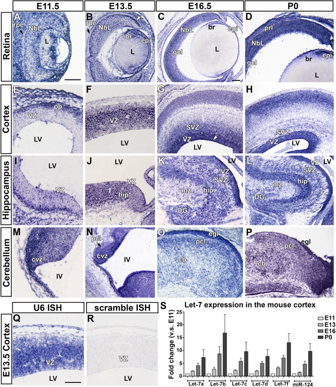
Let-7d expression in the embryonic mouse central nervous system. Let-7d expression pattern in the embryonic central nervous system analyzed using in situ hybridization and qRT-PCR. (A–D) Sagittal sections of the mouse retina at E11.5 (A), E13.5 (B), E16.5 (C) and P0 (D). Let-7d is absent from the retinal pigment epithelium (black arrowheads in A,B) and high along the apical surface (white arrowhead in B). (E–H) Horizontal sections of the mouse cerebral cortex at E11.5 (E), E13.5 (F), E16.5 (G) and P0 (H). From E16.5 onwards, the level of let-7d is particularly high along the apical surface of the lateral ventricle (white arrowhead in G). (I–L) Horizontal sections of the mouse hippocampus at E11.5 (I), E13.5 (J), E16.5 (K) and P0 (L). (M–P) Horizontal sections of the mouse cerebellum at E11.5 (M), E13.5 (N), E16.5 (O) and P0 (P). (Q,R) In situ hybridization using the following control probes: U6 positive control (Q) and scrambled negative control (R) on a horizontal section of the mouse cortex at E13.5. (S) qRT-PCR for let-7a,b,c,d,f and miR-124 on samples of mouse cortex collected at E11, E13, E16 and P0. All let-7 family members, and miR-124, increase over developmental time. Images shown in (A,B,E,F,I,J,M,N,Q,R) were taken at 20 × (scale bar in A = 100 µm). Images shown in (C,D,G,H,K,L,O,P) were taken at 10 × (scale bar in C = 100 µm). Rpe, retinal pigment epithelium; Nbl, neuroblastic layer; L, lens; br, bow region of the lens; epi, epithelium of the lens; gcl, ganglion cell layer; prl, photoreceptor layer; VZ, ventricular zone; LV, lateral ventricle; SVZ, subventricular zone; hip, hippocampus; mz, marginal zone; DG, dentate gyrus; CVZ, cerebellar ventricular zone; IV, fourth ventricle; pcl, Purkinje cell layer; cb; cerebellum; ecl, external germinal layer.

