Figure 3.
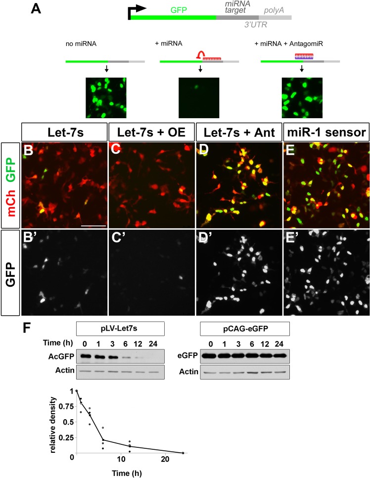
Let-7 is active in human embryonic retina-derived HER10 cells. (A) Schematic of a miRNA sensor. miRNA target sites are added to the 3’UTR end of GFP. If the miRNA of interest is present, GFP will be repressed (middle panel in A). In contrast, when no miRNA is present (left panel) or when a miRNA antagomiR is added (right panel), GFP is abundantly expressed. (B) GFP is present in 20–30−1.64 of HER10 cells transfected with pCAG-mCherry (red) and pLVX-let-7 sensor (let-7s). (C) Sensor-GFP expression is repressed when let-7 levels are increased via transfection of pLV-let-7 (OE; compare B’ to C’). (D) Co-transfection with a let-7 antagomiR (Ant) leads to GFP expression in almost all transfected cells. (E) Nearly all cells transfected with a miR-1 sensor (miR-1s) express GFP (E’). (F) Western blot analysis of a cyclohexamide time-course (time (h) = time after cyclohexamide addition) to determine the half-life of AcGFP1 present in the let-7s construct. The half-life of AcGFP1 is short, approximately 3.5 h (left and bottom panels), as compared to eGFP which was > 24 h (right panel). Unaltered images of Western Blots are included in the Supplementary Information (See Fig. S4).

