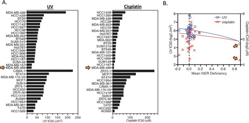
Figure 3:

UV and cisplatin sensitivity of breast epithelial and cancer cell lines. A. UV (left) and cisplatin (right) sensitivity is plotted for each mammary epithelial and breast cancer cell line. The MDA-MB-468 cell line (denoted by orange arrows) was the most sensitive cell line to both UV and cisplatin. B. UV and cisplatin IC50 values are plotted against mean NER deficiency (from the DDB2 proteo-probe assay) for each cell line. The MDA-MB-468 cell line (denoted by orange arrows) has the highest mean NER deficiency and lowest UV and cisplatin IC50 of all cell lines.
