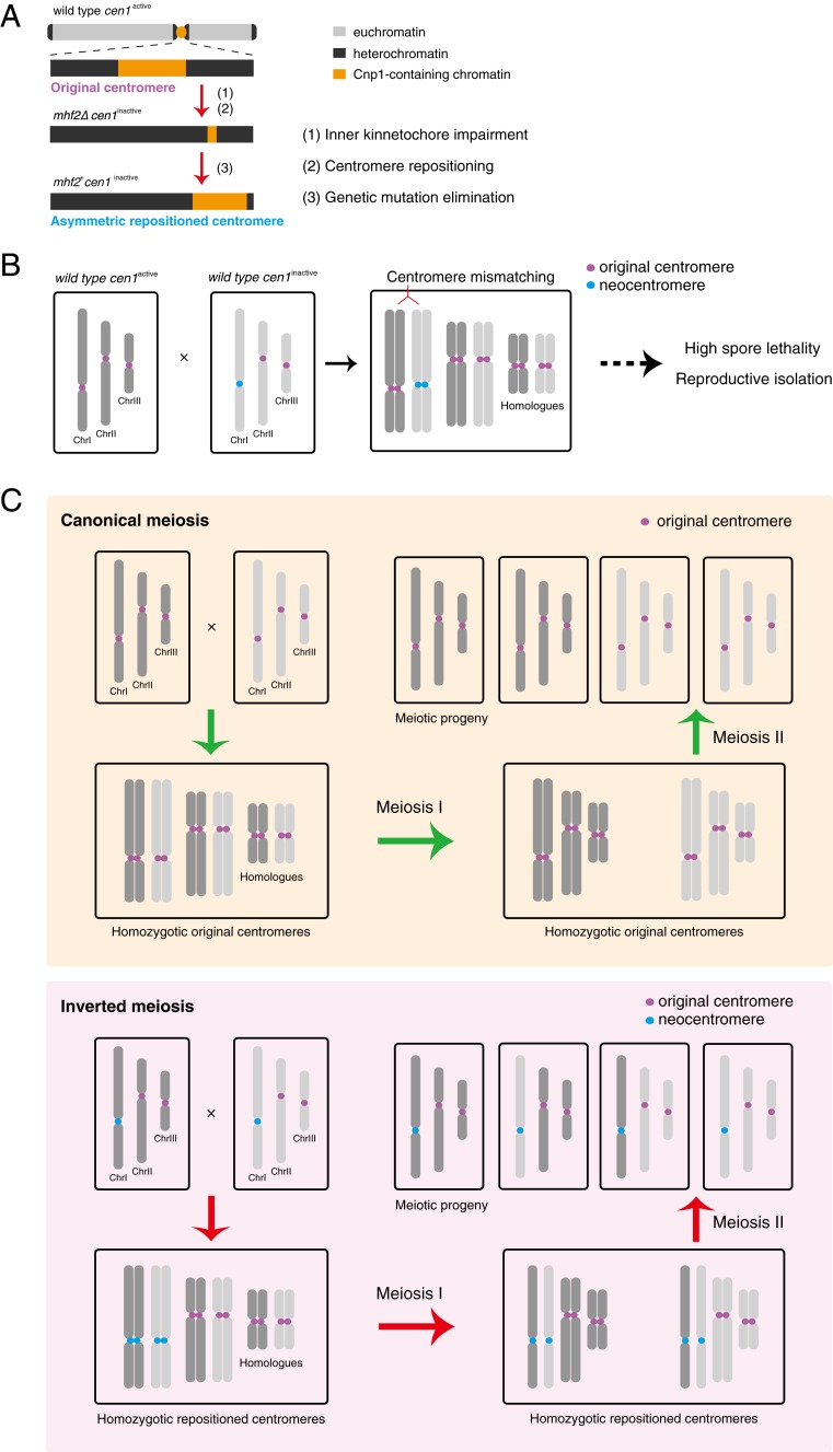
Fig. 8.
Model diagram of centromere repositioning. (A) Centromere inactivation is induced by impairment of the inner kinetochore, with subsequent asymmetric neocentromere formation preferentially at the pericentromeric regions. (B) Centromere mismatching between an original centromere (cen1active) and a neocentromere (cen1inactive) generates a reproductive barrier by causing severe spore lethality. (C) Canonical meiosis with homozygotic original centromeres (Upper) and inverted meiosis with homozygotic neocentromeres (Lower) both generate four viable meiotic progeny. In canonical meiosis, sister chromatids are cosegregated in first meiotic division (meiosis I) and are separated in the second meiotic division (meiosis II). In inverted meiosis, sister chromatids are disassociated from each other during meiosis I and the homologs are segregated during meiosis II. For simplicity, other features of meiosis including crossover, recombination between homologs and random segregation of homologs are omitted in this diagram.

