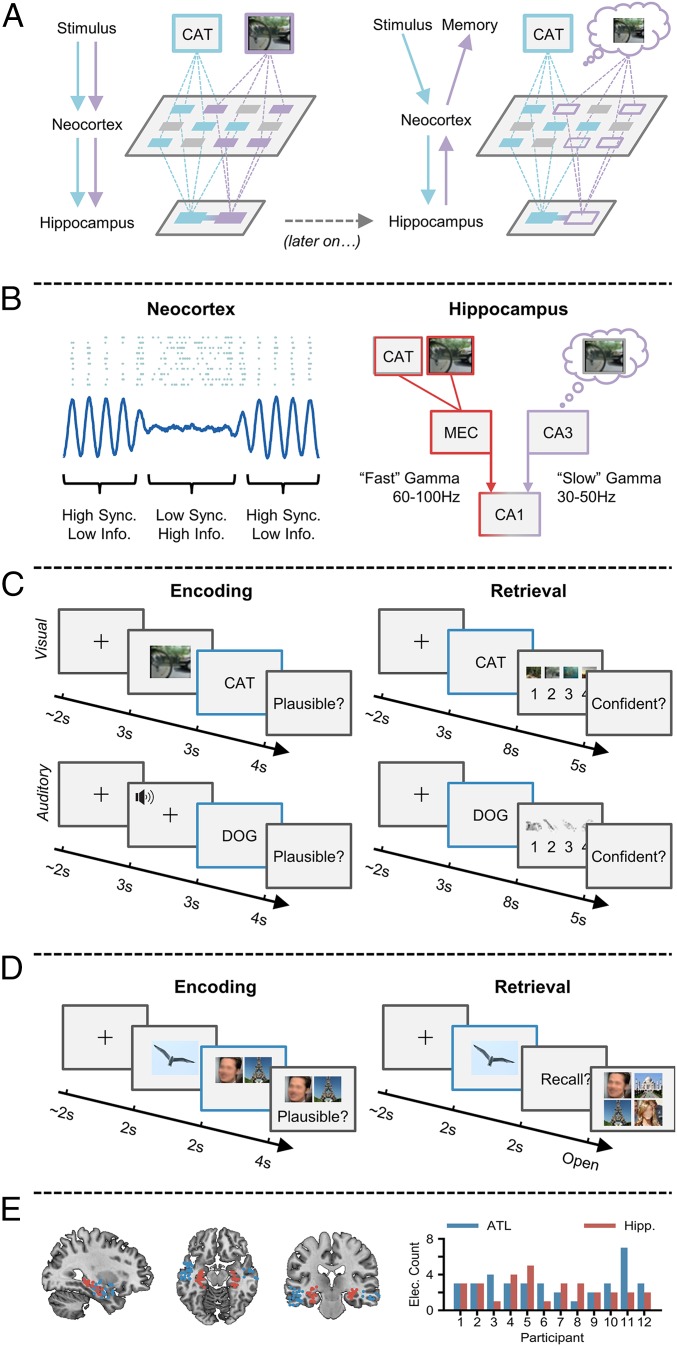
Fig. 1.
Synchronization–desynchronization framework. (A) Incoming stimuli are independently processed by relevant sensory regions of the neocortex (Left), and then passed on to the hippocampus where they are bound together. At a later stage (Right), a partial cue reactivates the hippocampal associative link, which in turn reactivates neocortical patterns coding for the memory representation, giving rise to conscious recollection. (B) Reduced oscillatory synchronization (blue line) within the neocortex allows individual neurons (blue dots) to fire more freely and create a more flexible neural code. Fast gamma activity allows the transfer of neocortical information to the hippocampus by boosting connectivity between the entorhinal cortex (MEC) and CA1. Slow gamma enhances retrieval by boosting connectivity between CA3 and CA1, allowing reinstated memories to be passed to the neocortex. (C) During encoding, participants are tasked with forming an associative link between a life-like dynamic stimulus (either a video or sound) and a subsequent verbal stimulus. During retrieval, participants are presented with verbal stimuli from the previous encoding block and asked to retrieve the associated dynamic stimulus. Electrophysiological analysis was conducted during the presentation of the verbal stimulus at encoding and retrieval (blue outline). (D) During encoding, participants are tasked with forming an associative link between an object, a face, and a scene. During retrieval, participants are presented with the object and asked to retrieve the associated face and scene. Electrophysiological analysis was conducted during the presentation of the verbal stimulus at encoding and retrieval (blue outline). (E) Plot of each electrode location (Left; red represents hippocampal electrode; blue represents the ATL). Bar plot (Right) depicts the number of electrodes for each participant.

