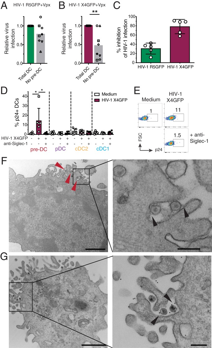
Fig. 2.
Pre-DC preferential infection is associated with Siglec-1–mediated capture of HIV-1. (A) Quantification of relative infection of total DCs (CD45+Lin(CD3/CD14/CD16/CD19/CD20/CD34)–HLA-DR+) or total DCs depleted of pre-DCs (CD33+CD45RAintCD123+Axl+) exposed to HIV-1 R5GFP + Vpx or (B) to HIV-1 X4GFP + Vpx. GFP expression was analyzed 48 h postinfection. (C) Relative quantification of the inhibition of the HIV-1 infection by preincubation with an anti–Siglec-1 mAb. Pre-DCs, preincubated with an anti–Siglec-1 mAb or not before, were infected for 48 h with HIV-1 X4GFP or R5GFP. Infection rates were quantified by following GFP expression. (D) Quantification of HIV-1 capture by the 4 DC subsets. Sorted DCs were preincubated for 30 min with an anti–Siglec-1 mAb or not, then exposed or not to HIV-1 X4GFP for 2 h at 37 °C, washed, stained for p24 to detect bound particles, and assayed by flow cytometry (HIV-1 X4GFP viral particles are not GFP+ by themselves), n = 5 or 6 independent donors combined in 5 experiments. Individual donors are displayed with bars representing mean ± SD. (E) Representative dot plot analysis of HIV-1 capture by pre-DCs performed as in D. (F) Ultrastrucutral analysis of pre-DCs exposed for 2 h to HIV-1 R5GFP, washed, and processed for EM. An epon section is presented, with a magnified view on the Right. Arrowheads point to virions captured by pre-DCs. On the magnified view, arrowheads point to protein linking the virions to the plasma membrane of the cells. (Scale bar, 2 µm [Left image] and 0.5 µm for the magnified view.) (G) Immuno-EM analysis of pre-DCs exposed to HIV-1 R5GFP for 2 h fixed and stained for Siglec-1 with a mAb revealed by protein A gold coupled to 5 nm gold particles before embedding (Materials and Methods). An epon section is presented, with a magnified view. Arrowheads point to Siglec-1–specific staining at the interface between virions and plasma membrane invaginations. (Scale bar, 2 µm [Left image] and 0.5 µm for the magnified view.) *P < 0.05, **P < 0.01.

