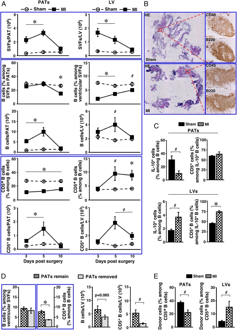
Fig. 6.
Impact of acute MI on the B cell compartment in PATs and LVs. Adult WT B6 mice were used. (A) SVFs from the indicated tissues of mice that underwent the indicated treatment were examined at the indicated time postsurgery. Summary of 2 to 3 independent experiments (n = 8 to 12 at each time point) for the indicated parameters are shown. (B) Sham- or MI-operated mice were examined 5 d postsurgery. Serial paraffin-embedded sections of PATs together with pericardium were either stained with H&E (Left, magnification: 0.5×) or immunostained for the indicated markers (Right, magnification: 20×). Representative graphs from 2 independent experiments are shown (n = 4). (C) Mice underwent the indicated surgery and were analyzed 7 d postsurgery. SVFs from the indicated tissues were cultured as described in the legend for Fig. 2B, in the presence of PIL. Cells were examined for IL-10+ B cells (summary of 2 independent experiments, n = 6). (D) Mice underwent the indicated procedures in conjunction with acute MI, and were examined 7 d later. Frequencies (Left) and total numbers (Right) of B cells and CD5+ B cells in LVs are shown (summary of 2 independent experiments, n = 6). (E) B cells from PerC of CD45.1+ mice were transferred into the pleural cavity of CD45.2+ mice during sham or MI surgery. Mice were examined 7 d later for donor cells among CD5+ B cells in the indicated tissues (summary of 3 independent experiments, n = 7). #P < 0.05 and *P < 0.01 for all panels.

