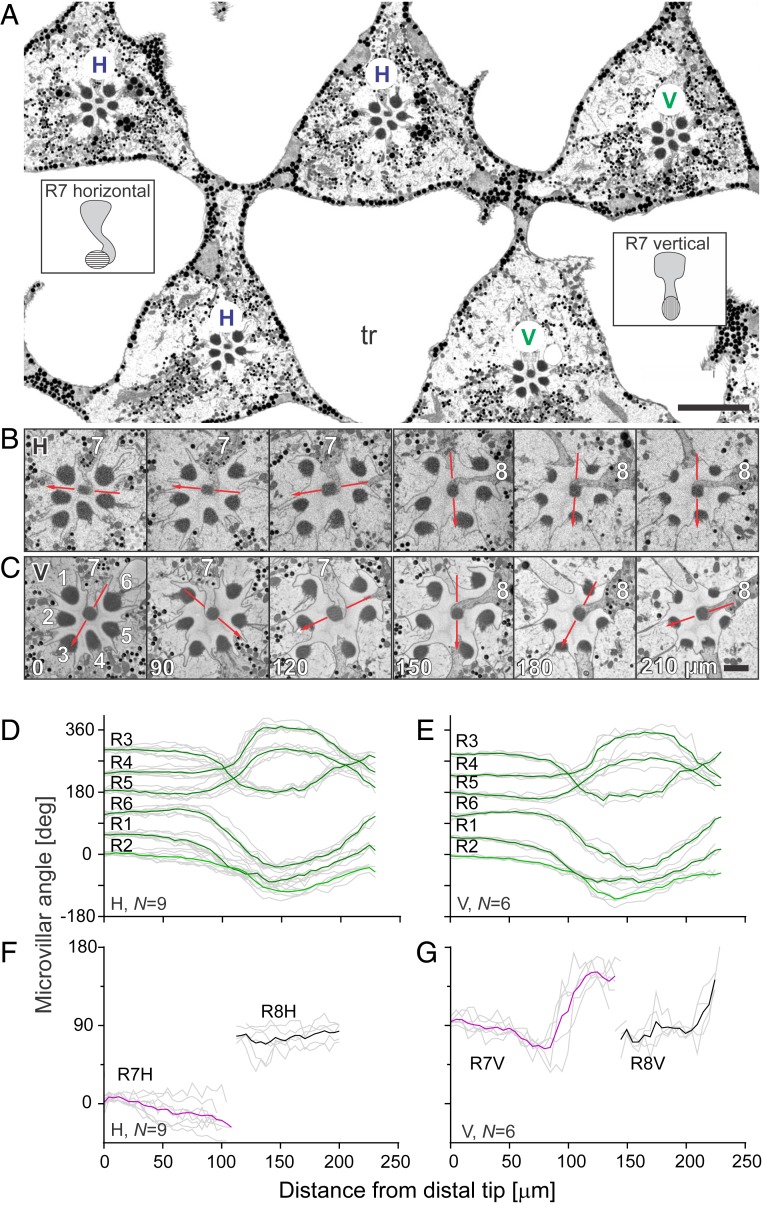
Fig. 1.
Anatomical analysis of rhabdomeric twisting in the frontal ventral retina. (A) Block surface scan of the distal retina with triangular ommatidia separated by large tracheoles (tr); the ommatidia are labeled according to the vertical (V) or horizontal (H) alignment of the microvilli in the central photoreceptor R7 (schematized in insets). (Scale bar, 10 µm.) (B and C) An H and a V ommatidium at depths, indicated in micrometers in C. Red lines indicate orientation of R7 and R8 microvilli. Photoreceptor identity is indicated with numbers. (Scale bars, 1 µm.) (D–G) Microvillar angle in rhabdomeres R1–6 (D and E) and R7 and R8 (F and G) as a function of the distance from the distal tip in the 15 analyzed ommatidia of type H (D and F) and type V (E and G). In D–G, colored and black lines indicate group means and gray lines individual rhabdomeres. N indicates the number of analyzed rhabdomeres.

