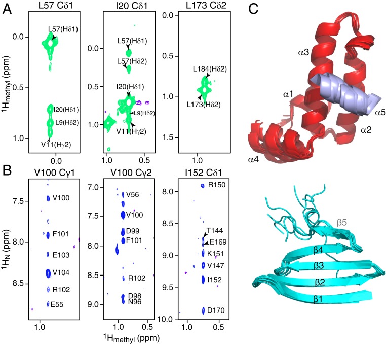
Fig. 3.
Methyl NOE data and domain structures of DNAJB6b. Strips from a 900-MHz 3D NOE-HMQC spectrum (250 ms mixing time) showing (A) methyl-methyl and (B) methyl-amide NOEs. The data were collected on a 200-μM [2H/15N/ILV-13CH3]-labeled ΔST-DNAJB6b sample at 25 °C. (C) Superposition of the 10 lowest-energy CS-ROSETTA structures calculated for the JD (Top; helices 1 to 4 in red and helix 5 in purple) and CTD (Bottom; cyan) domains. The flexible linkers are not shown for clarity.

