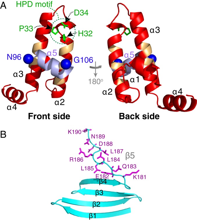
Fig. 5.
Structure of ΔST-DNAJB6b. Lowest-energy CS-ROSETTA structural model of the (A) JD and (B) CTD domains. In A, helix 5 is shown in purple, the nitroxide spin-label attachment positions are shown as blue spheres, the side chains of the HPD sequence motif are highlighted in green, and the backbone of residues previously identified to participate in the interface with Hsp70 (12) is indicated in orange. The side chains of the C-terminal 10 residues are shown in purple in B.

