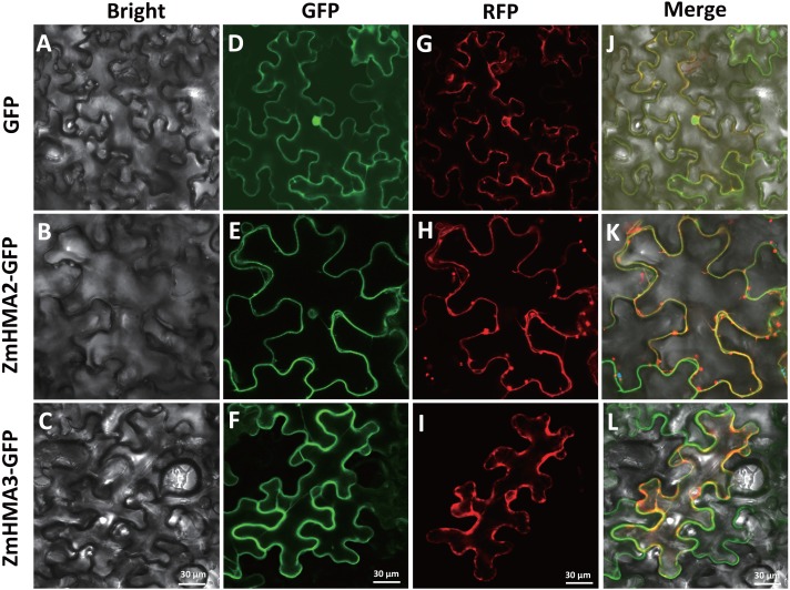
Figure 7. Subcellular localization of ZmHMA2 and ZmHMA3 in leaves.
(A–C) Tobacco cells under bright-field illumination. (D–F) Location of the fusion proteins, including the empty vector control, ZmHMA2 -GFP and ZmHMA3 -GFP under GFP-field. (G–I) show co-expression of a plasma marker (PIP2A-RFP), ZmHMA3 -GFP and ZmHMA3 -GFP under RFP-field. (J–L) Merged bright-field, GFP-field and RFP-field images. Scale Bars, 30 mm.

