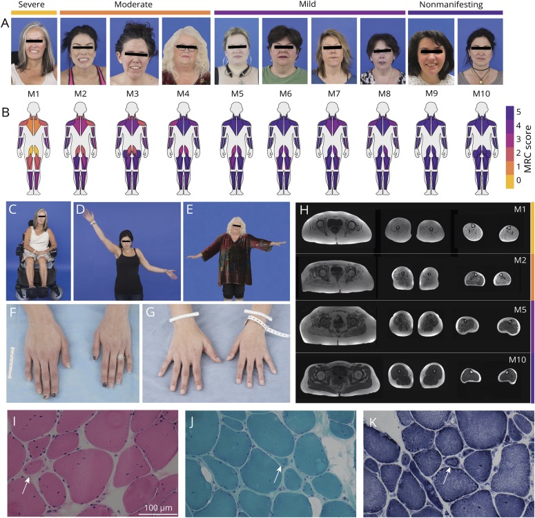
Figure 1. Clinical phenotype, muscle imaging findings, and muscle histotype.
(A) We phenotypically categorized carriers as severe (nonambulant) (M1), moderate (minimal independent ambulation/assisted ambulation) (M2–M4), mild (independent ambulation but with evidence of muscle weakness) (M5–M8), and nonmanifesting (no evidence of muscle weakness) (M9 and M10). (B) MuscleViz, a custom open-source software package, was created to visualize weakness using the Medical Research Council (MRC) scale for muscle strength. Plots can be generated using a free online browser application: muscleviz.github.io/. (C) The patient phenotypically categorized as most severe (M1) was wheelchair dependent by her third decade. (D, E) Patients M2 and M4's asymmetric weakness is revealed by the inability to raise the weaker arm as high as the stronger one. (F, G) Patients M2 and M3 have evidence of asymmetric skeletal growth, presenting as a difference in hand size, both with evidence of the left hand being smaller than the right hand. (H) Muscle MRI of the lower extremities reveals abnormal T1-weighted signal in all leg muscles imaged in M1 (severe), significantly increased T1 signal in the left medial gastrocnemius muscle, and normal T1 signal in the right medial gastrocnemius muscle in M2 (moderate) with prominent left–right asymmetry of lower leg muscle sizes below the knee, increased T1 signal in the right medial gastrocnemius muscle and right semitendinosus muscle in M5 (mild), and normal-appearing signal and size of all lower extremity muscles imaged in M10 (nonmanifesting). (I–K) Right vastus lateralis biopsy performed in M3 at 32 years of age with evidence of rare, subtle necklace fibers (arrows) on hematoxylin & eosin stain (×40) (I), Gömöri trichrome stain (×40) (J), and nicotinamide adenine dinucleotide stain (×40) (K). Measurement bar = 100 μm.

