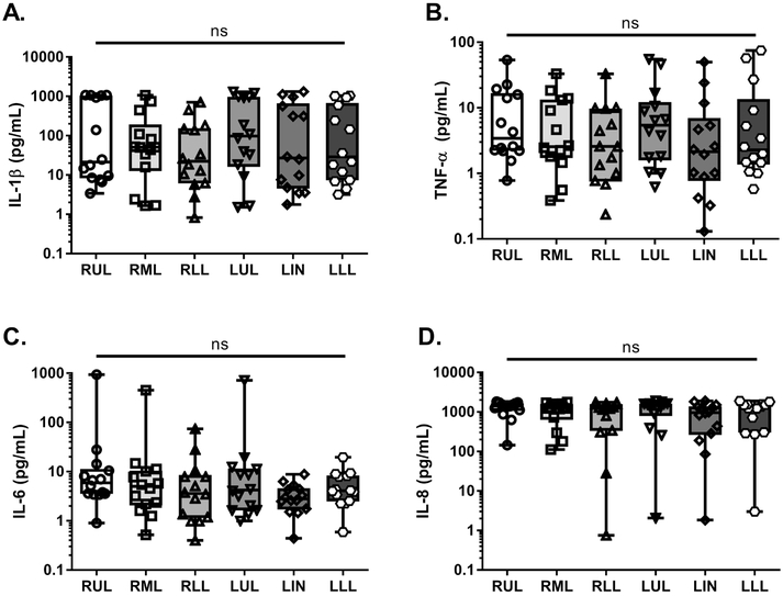
Fig. 2. Independent of infection, significant interlobar differences in proinflammatory cytokines were not observed.
Proinflammatory cytokine concentrations were measured in regional BAL specimens from all CF patients (n=14 patients= 84 lobar BALs) via V-PLEX array. Data from each specimen were stratified based upon lung lobe of origin. A. IL-1β (pg/mL) B. TNF-α (pg/mL) C. IL-6 (pg/mL) D. IL-8 (pg/mL). Statistical significance was determined via non-parametric, Kruskal–Wallis one-way analysis of variance (ANOVA). No statistically-significant differences were observed (ns). RUL- right upper lobe, RML- right middle lobe, RLL- right lower lobe, LUL- left upper lobe, LIN- lingula, LLL- left lower lobe.

