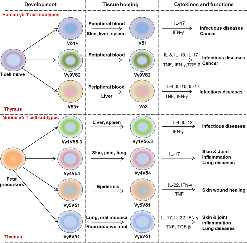
Figure 1: Overview of γδ T cell functional programing in human and mouse.
Schematic illustration of γδ T cell development from naïve fetal precursors in human (upper panel) and mouse (lower panel) thymus. Human naïve γδ T cells differentiate into Vδ1+ γδ T subtypes found in peripheral blood, skin, liver, and spleen and produce IL-17 and IFN-γ; Vγ9+Vδ2+ γδ T cells found in the peripheral blood and produce predominantly TNF and IFN-γ as well as IL-8, IL-10, IL-17; Vδ3+ found in peripheral blood and liver and produce several cytokines such as IL-4, IL-10, IL-17, TNF, and IFN-γ. In mouse, Vγ1+Vδ6.3+ γδ T cells normally resident in lymphoid tissues including spleen and liver and produce IL-4, IL-13, and IFN-γ. Vγ4+Vδ4+ γδ T cells are found in the skin, joint, and lung and known as IL-17-producing γδ T cells. Vγ5+Vδ1+ are residents in epidermis, produce IL-17, TNF, IFN-γ whereas Vγ6+Vδ1+ have been found in the female reproductive tract and oral mucosa and produce IL-17, IL-22, TNF, IFN-γ and TGF-β.

