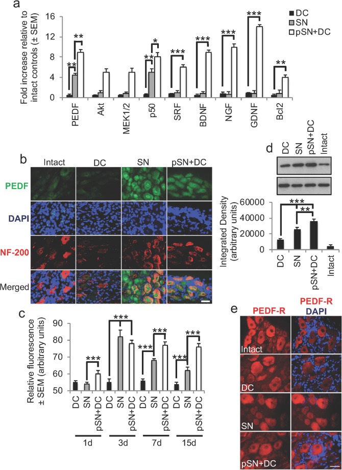
Fig. 2.
Confirmation of highly changed genes in pSN + DC DRGN by qRT-PCR and confirmation of changes in PEDF. a Confirmation of highly upregulated levels of PEDF, Akt, MEK1/2, p50, SRF, BDNF, NGF, GDNF, and Bcl2 by qRT-PCR. b Immunohistochemistry at 10 days after DC injury and c quantification of immunopositive staining at 1, 3, 7, and 15 days post-DC injury showed high levels of PEDF immunoreactivity (green) in regenerating SN and pSN + DC DRGN soma (red) (DAPI = blue, cell nuclei), whilst low levels were detected in intact and DC-injured DRGN. d Western blot and subsequent densitometry detected high levels of PEDF in regenerating SN and pSN + DC models, with the highest levels of PEDF being present in pSN + DC DRG. e PEDF-R (green) was present in DRGN and remained unchanged in intact and all injury models. DAPI (blue) = cell nuclei. Scale bars in b and e = 25 μm. * = P<0.05, ** = P < 0.001, ANOVA; *** = P < 0.0001, ANOVA

