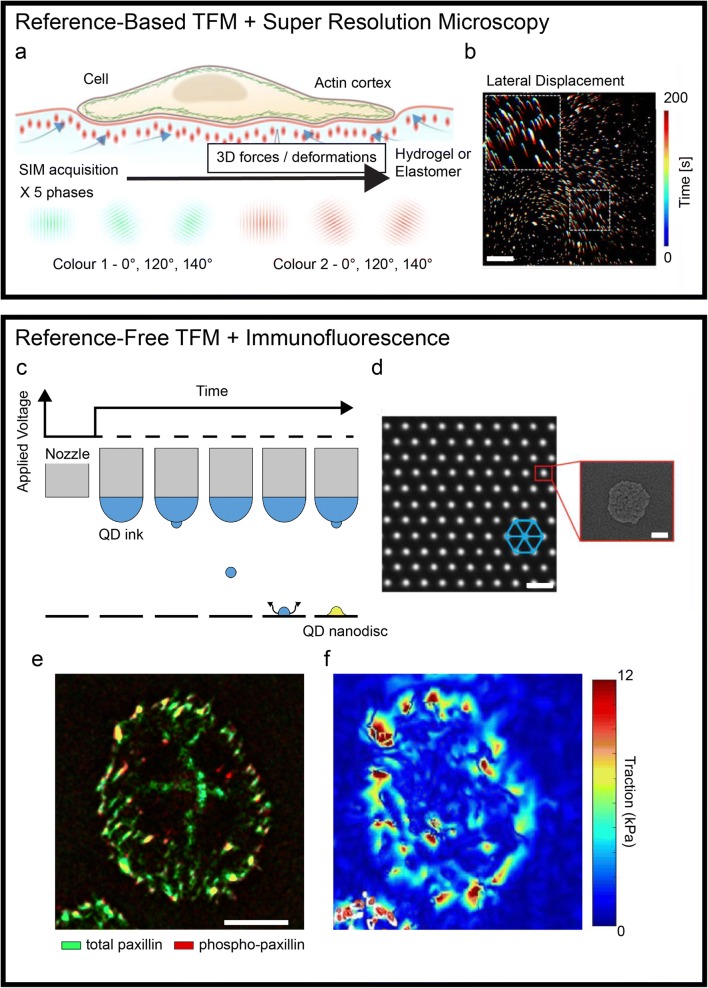
Fig. 1.
Recent advancements in traction force microscopy. A super-resolution microscopy applied to reference-based TFM substrates (top box). (a) Schematic representation of a structural illumination microscopy (SIM) applied to a compliant TFM substrate. (b) A temporal projection of fluorescent optical landmarks tracked by SIM, rendering the cell-induced displacement upon force generation. Scale bar is 5 μm. Printing technologies for reference-free TFM enabling the combination with immunofluorescence (bottom box). (c) Schematic of electrohydrodynamic NanoDrip printing. Induction of a liquid meniscus and ejection of ink nanodroplets upon application of DC voltage. Once impacted on the underlying substrate, the droplets are vaporized leaving a QD nanodisc (d, inset). The process is reiterated accurately moving the nozzle to obtain a triangular array of fiducial markers (d). Scale bar is 2 μm. (e–d) The resulting ordered placement allows the endpoint staining of cellular components. Maps of focal adhesion markers distribution can be directly overlapped to the corresponding tractions. Scale bar is 10 μm. Adapted from (Colin-York et al. 2019; Galliker et al. 2012; Bergert et al. 2016; Panagiotakopoulou et al. 2018)

