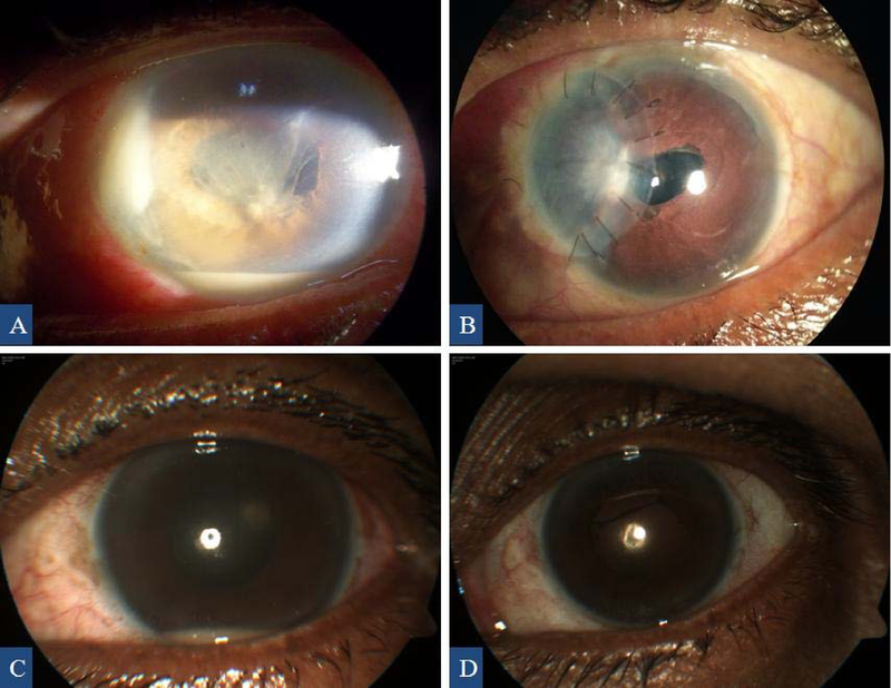
Figure 1.

Anterior segment images showing nasal corneal abscess, hypopyon, and fibrinous exudates at the time of presentation (A) and corneal patch graft at the last visit (B) of case 12. Anterior segment images of case 13 showing recurrence of hypopyon (C) and postoperative image at the last visit with no hypopyon (D).
