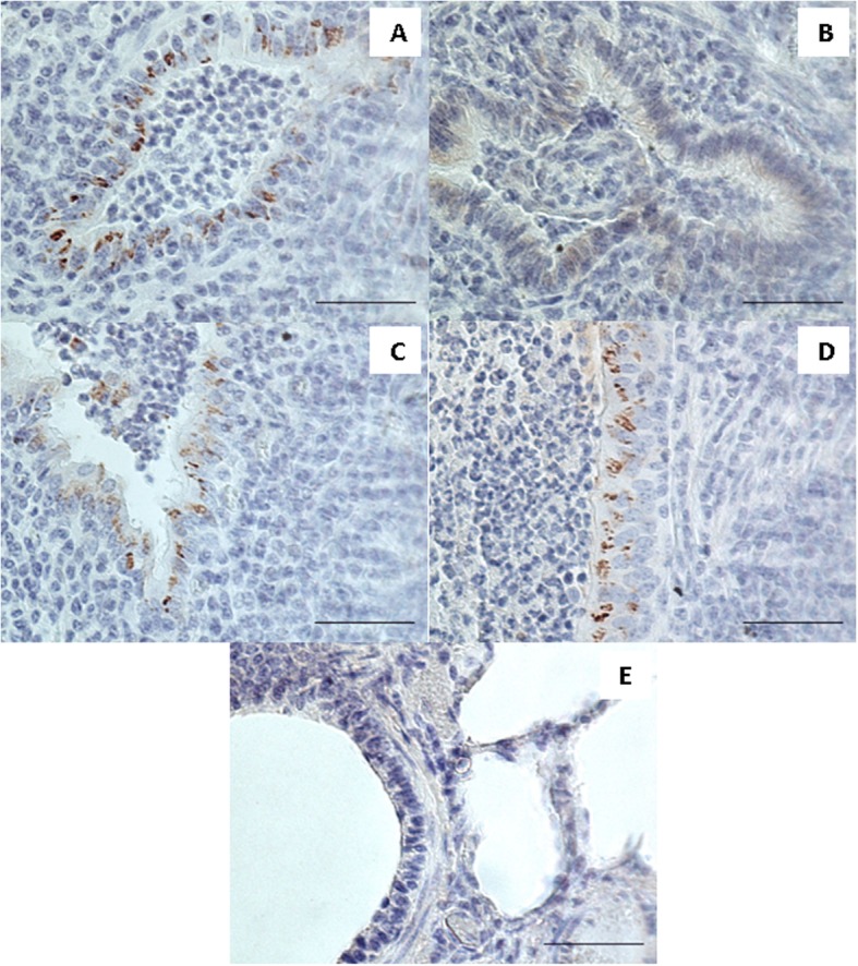
Fig. 1.

Immunohistochemistry. Positive labelling for M. bovis visible as dark brown granular aggregates was observed in epithelial cells of bronchioli in the lung of the positive control calf infected with M. bovis (a), the lung of the calf infected with M. bovis and received enrofloxacin combined with flunixin meglumine (c) and the lung of the calf infected with M. bovis that received enrofloxacin combined with flunixin meglumine and pegbovigrastim (d). The lung of the calf infected with M. bovis and received enrofloxacin alone showing lack of specific granular immunolabelling with faint diffuse light brown staining visible in bronchiolar epithelium (b). The lung of a negative control calf showing lack of labelling for M. bovis (e). Bar = 50 μm
