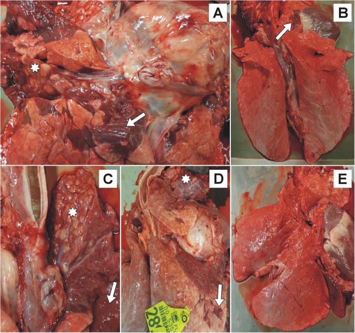
Fig. 5.
The lungs of treated and control calves. The lung of a positive control calf infected with Mycoplasma bovis (a): extensive caseous necrosis (star) and marble-like lesions (lobular consolidation, arrow). Lung from a calf infected with M. bovis and received enrofloxacin alone (b): a slight lobular consolidation (arrow). The lung of the calf infected with M. bovis and received enrofloxacin combined with flunixin meglumine (c): extensive caseous necrosis (star) and marble-like lesions (arrow). The lung of a calf infected with M. bovis that received enrofloxacin combined with flunixin meglumine and pegbovigrastim (d): extensive caseous necrosis (star) and marble-like lesions (arrow). The lung of a negative control calf (e): no lesions

