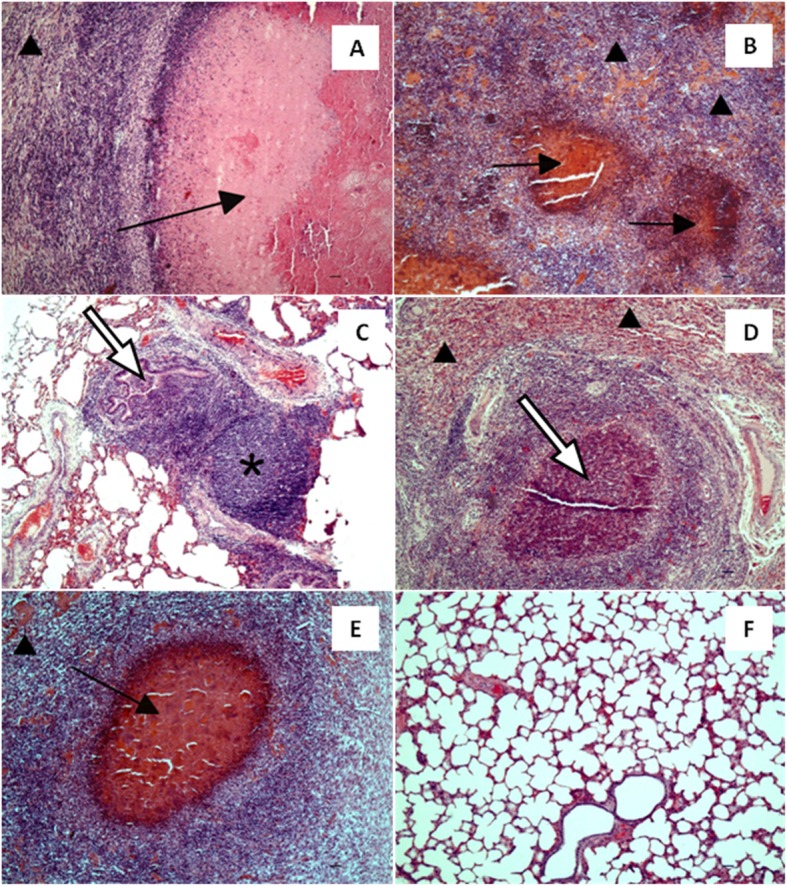
Fig. 6.

The lungs of treated and control calves. The lung of a positive control calf infected with Mycoplasma bovis (a): extensive caseous necrosis with amorphic eosinophilic core surrounded by macrophages and lymphocytes infiltrating surrounding lung parenchyma (black arrow); prominent atelectasia (arrow head). (b): multifocal inflammatory infiltrations with necrotic on centres containing macrophages and neutrophils, surrounded by lymphoidal cells (black arrow); prominent atelectasia (arrow head). The lung of the calf infected with M. bovis and received enrofloxacin alone (c): mild hyperplasia of bronchiolar-associated lymphoid tissue (star), as well as accumulation of neutrophils and macrophages in bronchiolar lumens (white arrow). The lung of a calf infected with M. bovis and received enrofloxacin combined with flunixin meglumine (d): extensive diffuse infiltration of neutrophils and macrophages and occasional foci of necrotic cells surrounded by macrophages and lymphoid cells (white arrow); prominent atelectasia (arrow head). The lung of the calf infected with M. bovis that received enrofloxacin combined with flunixin meglumine and pegbovigrastim (e): focal caseous necrosis (black arrow); prominent atelectasia (arrow head). The lung of a negative control calf (f). HE, Bar = 50 μm
