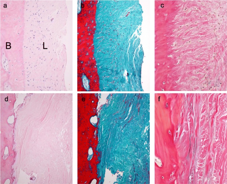
Fig. 7.

Images showing the histomorphometry using hematoxylin-eosin (left), Masson trichrome (middle) and Elastica van Gieson (right) staining of the distal attachment of the sMCL in the normal control a, b, 100x magnified, c 400x magnified. After removal of a locking plate d, e, 100x magnified, e 400x magnified. L: ligament B: bone
