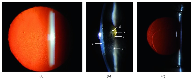
Figure 1.
Slit lamp biomicroscopy photos pre- and post-DSO surgery for the left eye. (a) Preoperative red reflex photo with a dilated pupil shows confluent guttae in the visual axis, slightly greater in the inferior half of the visual axis compared to superior half. The peripheral cornea is relatively spared of guttae. (b) Slit photo through the central cornea at 1 week post-DSO surgery demonstrates a. the edge of the descemetorhexis; b. guttae outside the area of Descemet's stripping; c. reticular epithelial edema; d. peripheral ring of corneal clearing without edema in presumed area of endothelial healing; and e. corneal stromal edema. (c) One month postoperative red reflex photo shows inferocentral descemetorhexis with clear visual axis without guttae and peripheral nonconfluent guttae.

