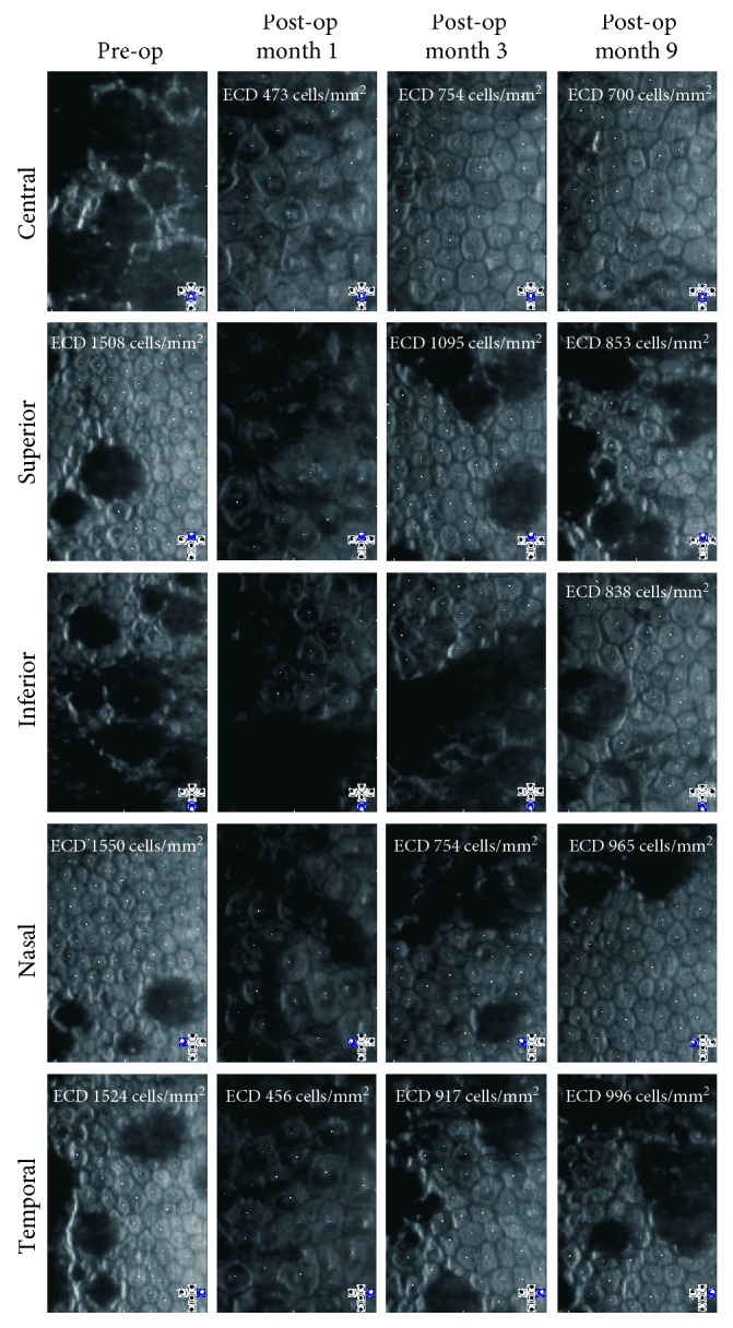
Figure 2.

Specular microscopy imaging of the corneal endothelium pre- and post-DSO surgery for the left eye. The locations (central, superior, inferior, nasal, and temporal) are based upon the standard gaze positions of the Konan specular microscope. Endothelial cell density (ECD) is indicated when measurable.
