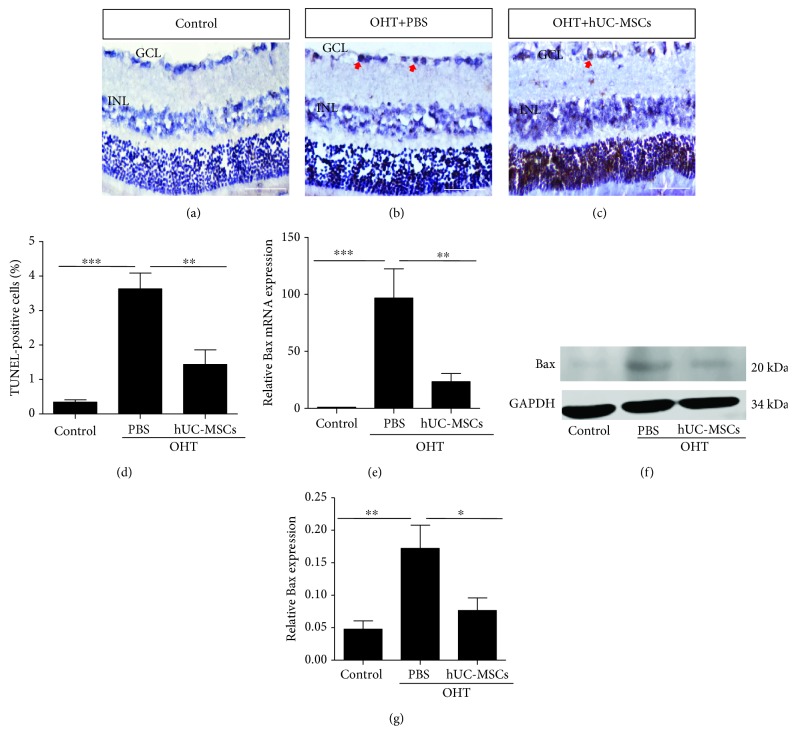
Figure 3.
hUC-MSC transplantation decreases the apoptosis of retinal cells in OHT-induced rats. Two weeks after hUC-MSC injection. (a–c) Representative images of TUNEL staining in retinas from normal eyes, OHT+PBS eyes, and OHT+hUC-MSC eyes. The positive cells in the GCL and ILN of retinas were brown, and the arrow indicates positive TUNEL staining. (d) Quantification of TUNEL-positive cells (n = 4/group; ∗∗P < 0.01 and ∗∗∗P < 0.001 compared with one-way ANOVA with Turkey's post hoc tests). (e) Relative Bax gene expression that was normalized to GAPDH in retinas from normal eyes, OHT+PBS eyes, and OHT+hUC-MSC eyes (n = 4/group; ∗∗P < 0.01 and ∗∗∗P < 0.001 compared with one-way ANOVA with Turkey's post hoc tests). (f) Western blot analysis for Bax protein from normal eyes, OHT+PBS eyes, and OHT+hUC-MSC eyes. (g) Relative Bax protein expression that was normalized to GAPDH (n = 4/group; ∗P < 0.05 and ∗∗P < 0.01 compared with one-way ANOVA with Turkey's post hoc tests). Scale bar: 100 μm.

