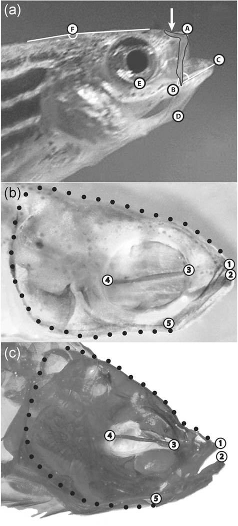
FIGURE 1.
Landmarks and semilandmarks used in analyses of movement and shape. (a) Landmarks used in kinematic analyses with an image of the premaxillary bone of the upper jawsuperimposed its correct anatomical position in a fully protruded upper jaw. Arrow indicates the ascending arm of the premaxilla. Landmarks: (A) Anterior tip of the upper jaw; (B) corner of the mouth; (C) anterior tip of the lower jaw; (D) anterior tip of the hyoid; (E) ventral‐most point of the orbit; and (F) vertex of the angle used to measure head rotation during cranial elevation (the dorsal surface of the head anterior to this point rotated upward during cranial elevation when feeding, while the dorsal surface of the trunk posterior to this point did not). (b) Anatomical landmarks and semilandmarks used in shape analyses of all specimens at all ages sampled shown on a larval zebrafish. Landmarks: (1) anterior tip of the premaxilla in the upper jaw; (2) anterior tip of the dentary bone in the lower jaw; (3) junction of the parasphenoid with the anterior wall of the orbit; (4) junction of the parasphenoid with the posterior wall of the orbit; and (5) lower jaw joint (articularquadrate joint). Black circles indicate semi‐landmarks evenly spaced between LM 1 and 2 to capture overall head shape. (c) Anatomical landmarks and semi‐landmarks used in shape analyses of all specimens at all ages sampled shown on an adult zebrafish (LM are the same as those in panel b). LM, landmark

