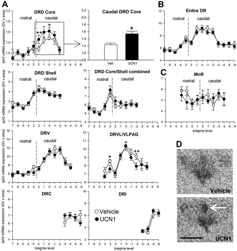
Figure 2.
Effects of five days of daily bilateral intra-bed nucleus of the stria terminalis (BNST) microinjections of vehicle (n = 11, open circles) or urocortin 1 (UCN1, n = 11, closed circles) on tph2 mRNA expression in subdivisions of (A) the dorsal raphe nucleus (DR), (B) in the entire DR (average of all subdivisions), and (C) in the median raphe nucleus (MnR). Tph2 mRNA expression is displayed throughout the entire rostrocaudal extent of each subdivision, with bregma levels (+7 through −6) on the x-axis, and gene expression on the y-axis. Borders between rostral and caudal aspects of each subdivision are indicated with dashed vertical lines. Shown is the mean ± or + the standard error of the mean (SEM). (D) Representative photomicrographs showing tph2 mRNA expression in the DR of vehicle- and Ucn1-primed rats at approximately −8.168 mm from bregma. The white arrow indicates the core of the caudal aspect of the dorsal raphe nucleus, dorsal part(cDRD core). Abbreviations: DRC, dorsal raphe nucleus, caudal part; DRD, dorsal raphe nucleus, dorsal part; DRI, dorsal raphe nucleus, interfascicular part; DRV, dorsal raphe nucleus, ventral part; DRVL/VLPAG, dorsal raphe nucleus, ventrolateral part/ventrolateral periaqueductal gray; Scale bar: 1 mm. p* < 0.05, p** <0.01, compared to vehicle controls.

