Fig. 4.
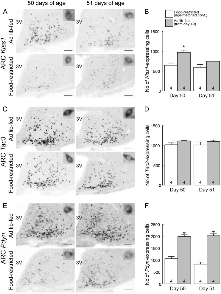
Ad libitum feeding increased the number of Kiss1- and Pdyn-expressing cells in the arcuate nucleus (ARC) of the growth-retarded female rats. (A) Kiss1-expressing cells in the ARC of representative female rats subjected to ad libitum feeding on 50 and 51 days of age (24 and 48 h after the resumption of ad libitum feeding) and age-matched food-restricted controls. (B) The number of Kiss1-expressing cells throughout the ARC of ad libitum-fed rats on 50 and 51 days of age and age-matched food-restricted controls. (C) Tac3-expressing cells in the ARC of representative female rats subjected to ad libitum feeding on 50 and 51 days of age and age-matched food-restricted controls. (D) The number of Tac3-expressing cells throughout the ARC of ad libitum-fed rats on 50 and 51 days of age and age-matched food-restricted controls. (E) Pdyn-expressing cells in the ARC of representative female rats subjected to ad libitum feeding on 50 and 51 days of age and age-matched food-restricted controls. (F) The number of Pdyn-expressing cells throughout the ARC of ad libitum-fed rats on 50 and 51 days of age and age-matched food-restricted controls. Insets show representative cells showing Kiss1 (A), Tac3 (C) or Pdyn (E) expression at higher magnification. Scale bars, 100 μm; 3V, third ventricle. Values are expressed as the mean ± SEM. Numbers in each column indicate the numbers of animals used. Asterisks indicate significant difference in the number of ARC Kiss1- or Pdyn-expressing cells between the ad libitum-fed rats and the age-matched food-restricted controls (* P < 0.05, Student’s t-test).

