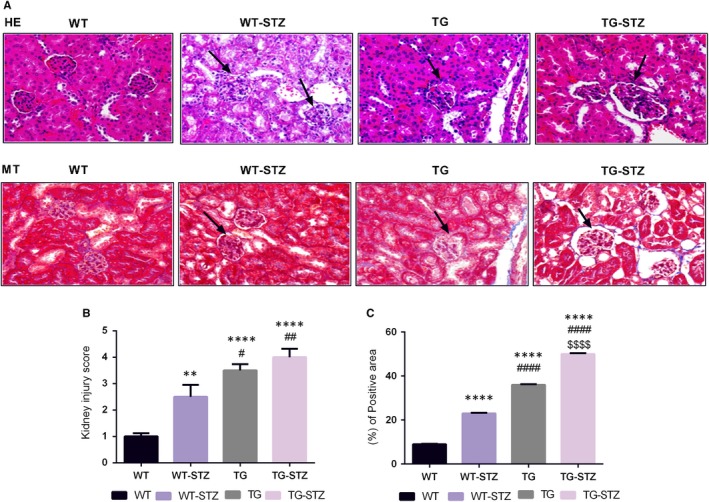
Figure 1.

Histological examinations of kidney tissues from WT, WT‐STZ, TG and TG‐STZ (A). Haematoxylin and eosin staining and Masson's trichrome staining (B). Kidney injury score of four groups (C). Collagen deposition was higher in TG‐STZ. **P < .01 are compared to the WT; ****P < .0001 are compared to the WT; #P < .05 are compared to the WT‐STZ; ##P < .01 are compared to the WT‐STZ; ####P < .0001 are compared to the WT‐STZ; and $$$$P < .0001 are compared to the TG
