
Figure 2.
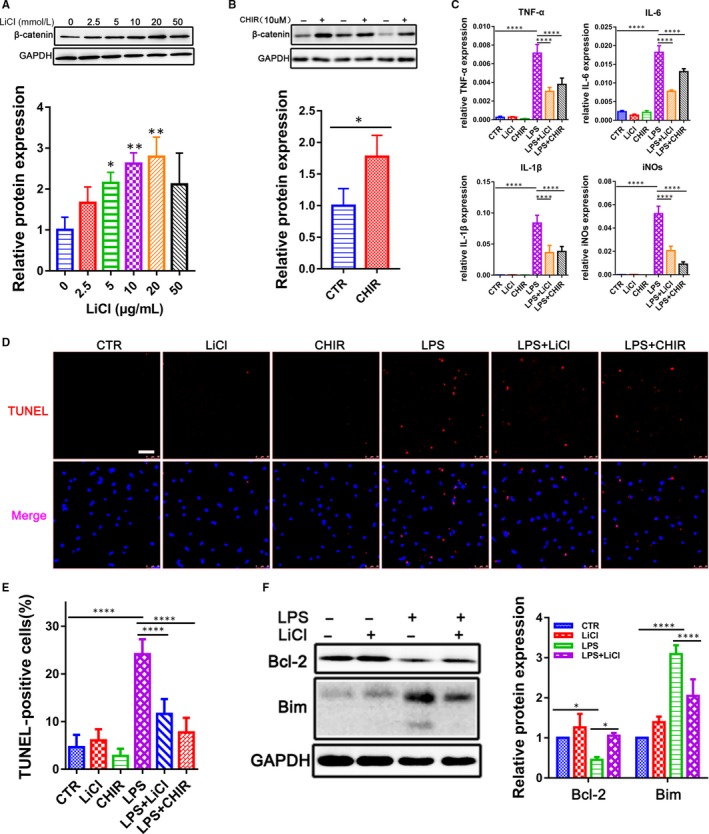
Down‐regulation of GSK‐3β attenuates myocardial inflammatory injury in cardiomyocytes after LPS challenge. A, B, Relative β‐catenin expression was measured by Western blot in the CMs treated with GSK‐3β inhibitors LiCl (0‐50 mM) and CHIR‐99021 (10 μM) for 24 h (n = 3). C, qRT‐PCR analysis for pro‐inflammatory cytokines TNF‐α, IL‐1β, IL‐6 and iNOs in CMs treated with LiCl (10 mM) and CHIR‐99021 (10 μM) in the presence of LPS (500 ng/mL) for 12 h (n = 3). D, E, TUNEL assay for apoptosis in CMs treated with LiCl (10 mM) and CHIR‐99021 (10 μM) in the presence of LPS (500 ng/mL) for 12 h. Nuclei were counterstained with the DNA‐intercalating dye Hoechst (blue). The lower panel was the percentage of TUNEL‐positive cells (n = 3). (Scale bar: 25 μm). F, Western blot for Bim and Bcl‐2 in CMs pre‐treated with LiCl (10 mM) for 12 h and followed by stimulation of LPS (500 ng/mL) for another 12 h. GAPDH was loaded as internal control (n = 3). *P < .05; **P < .01 and ****P < .0001 when compared with controls