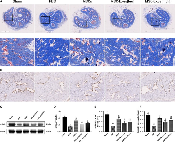
Figure 6.

Treatment improves the ratio of smooth muscle to collagen and smooth muscle content in the corpus cavernosum. A, The smooth muscle (red) and collagen (blue) tissues stained by Masson's trichrome staining. Original magnification, ×20 and × 100. B, Immunohistochemical expression of α‐SMA in corpus cavernosum. Original magnification, ×100. C, Representative images of western blots for α‐SMA in cavernosum in each group. D, Western blots' data are presented as the relative density of α‐SMA compared with that of GAPDH. E, Immunohistochemically semi‐quantitative data of the proportion of α‐SMA positive expression area. F, The semi‐quantitative data of the ratio of smooth muscle area to collagen area. Each bar depicts the means ± standard deviation from n = 6 animals per group. *P < .05 vs the PBS group, **P < .01 vs the PBS group, #P < .05 vs the Sham group, ##P < .01 vs the Sham group
