Figure 1.

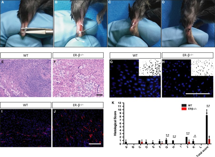
ERβ−/− tendon scars have an inferior tendon repair process. A‐D, Surgery process of mouse Achilles tendon. E, F Low‐magnification HE staining indicates a very different scar organization with clear adipocyte accumulation in ERβ−/− mice. (G,H) Cell density in the healing region was significantly lower in ERβ−/− mice compared with WT controls. (I,J) Number of CD34‐labelled blood vessels was significantly higher in ERβ−/− mice compared with WT controls. K, Evaluation of tendon healing using an established histological scoring system revealed that ERβ−/− mice had a significantly lower histological score compared with WT controls. Data are represented as mean ± SEM (n = 5), *P < .05, **P < .01. Scale Bars: 100μm
