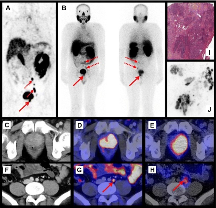
Figure 2.
Preoperative imaging using 68Ga-PSMA-11 PET/CT 1 h p.i. (A) and 111In-PSMA-I&T SPECT/CT and planar scintigraphy (4 h p.i., 155 MBq) (B). Axial 68Ga-PSMA-11 PET/CT images of the primary tumor in the prostate (D) and a representative lymph node (G). Corresponding CT images (C, F) and axial 111In-PSMA-I&T SPECT/CT images (E, H). H&E staining (I) and 111In-autoradiography (J) of cryosections from resected prostate tissue. The human study was approved by the institutional review boards of the participating medical institutions, and the patient provided signed informed consent. Reprinted with permission from Schottelius et al., 111In-PSMA-I&T: expanding the spectrum of PSMA-I&T applications towards SPECT and radioguided surgery, Copyright 2015, Springer 22.

