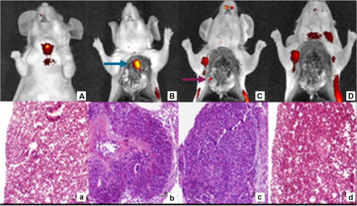
Figure 4.
Sequential tumor debulking surgery and H&E analysis of 22RV1 tumor metastases 4 h p.i. with DUPA-IRDye800CW (10 nmol). Fluorescent and white light image overlays of whole body image (A), opened chest cavity (B), after the removal of the primary tumor (blue arrow) (C) and after the removal of all secondary nodules (purple arrow) (D). H&E staining healthy control lung (a), primary tumor (b), secondary tumor nodule (c) and residual tissue (d). Reprinted with permission from Kelderhouse et al., Development of tumor-targeted near infrared probes for fluorescence guided surgery, Copyright 2013, American Chemical Society 52.

