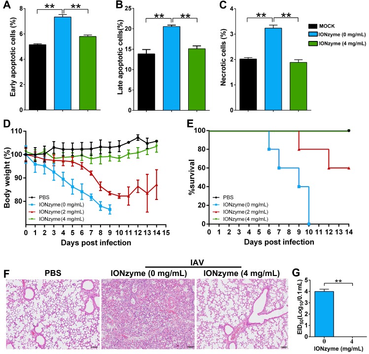
Figure 5.
IONzymes reduce IAVs-induced apoptosis in vitro and pathogenicity in vivo. (A-C) Apoptosis analysis of MDCK cells infected with IONzyme-treated IAVs (H5N1 SY strain) using flow cytometry. MOI = 1 under 24 h p.i.. The proportions of early apoptotic cells (annexin V+ and PI-; A), later apoptotic cells (annexin V+ and PI+; B), and necrotic cells (annexin V- and PI+; C) were analyzed. (D-G) BALB/c mice were challenged intranasally with IONzyme-treated of IAVs. (D) Morbidity was evaluated by monitoring weight changes over a 14-day period and was plotted as a percentage of the animals' weights on the day of inoculation (day 0). The data represent the average body weight of each group (n = 5). (E) Mortality expressed as percent survival. (F) Representative histopathological changes in H&E (hematoxylin and eosin)-stained lung tissues on day 7 p.i.. (G) Viral replication in the lung (n = 3). Data shown represent the means±s.d. of three samples. Statistical significance is assessed by unpaired Student's two-sided t-test (two groups). **P<0.01. Scale bars: 500 μm.

