Figure 1.
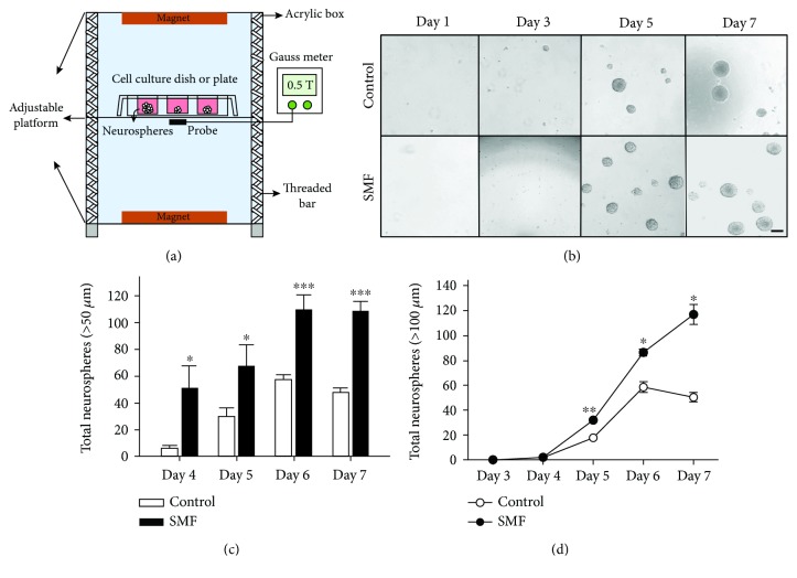
SMF stimulates the proliferation of mNPCs. Neurospheres developed from the neurosphere assay of control and SMF group. (a) Schematic drawing of the experimental device. (b) Typical phase-contrast micrographs. Scale bar, 100 μm. (c) Neurosphere numbers per well developed from 4, 5, 6, and 7 day cultures. The number of neurospheres is significantly increased under SMF exposed. (d) Neurosphere numbers per well at sizes (>100 μm) developed from 3-7 day cultures. Data represent the mean ± S.E.M.; control, n = 5; SMF, n = 3; ∗p < 0.05, ∗∗p < 0.01, ∗∗∗p < 0.001; Student's t-test.

