FIG. 1.
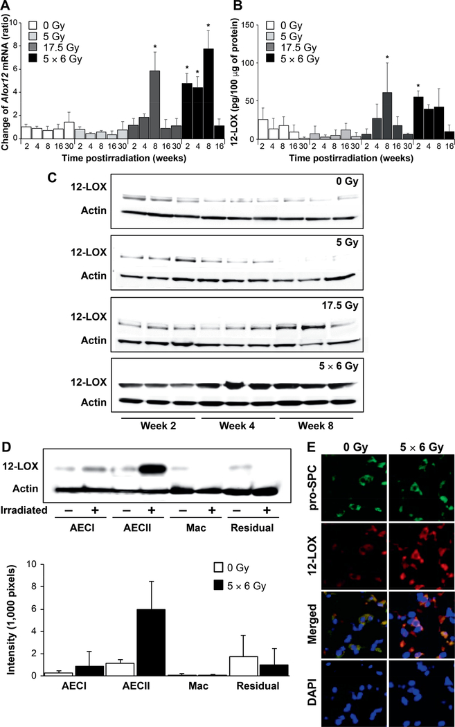
Increased expression of 12-LOX in irradiated lungs. C57/Bl6NcR mice, 8–10 weeks old, received thoracic irradiation (0 Gy, 5 Gy, 17.5 Gy or 5 × 6 Gy) and maintained for 2, 4, 6, 8, 16 or 30 weeks. Lung tissue was collected at the indicated timepoints. Panel A: RNA was isolated from lung tissue at expsoure and timepoints noted (n = 3 mice per condition). The expression of Alox12 was determined using RT-PCR and normalized to β-actin. The normalized expression of Alox12 is presented relative to 0 Gy, week 2. Panel B: The concentration of 12-LOX in homogenized lung tissue was determined with ELISA at the indicated timepoints postirradiation. Panel C: Protein expression of 12-LOX in irradiated lung tissue was confirmed by Western blotting of lung tissue lysates. Panel D: Lung tissue from mice that received thoracic irradiation at varying doses was digested to a single cell suspension and macrophage, pneumocyte and remaining cell populations were enriched for analysis. The expression of 12-LOX was determined in protein lysates from enriched cell populations (n = 3); representative blot shown. Densitometry was used to quantitatively represent differences in 12-LOX expression relative to actin. Panel E: Lung tissue collected at 8 weeks postirradiation was subjected to immunofluorescence to localize 12-LOX expression relative to that of prosurfactant-C, a marker of type 2 pneumocytes. Columns are the mean and bars represent standard deviation (SD), *P < 0.05 compared to 0 Gy, week 2 by ANOVA. Mac = macrophages; residual = residual digested lung cellular content after AECI, AECII and macrophages removed. pro-SPC = pro-surfactant C.

