
Table 1.
Prototypical small-molecule Sigma1 and Sigma2/TMEM97 modulators/ligands.
| Compound | Binding affinity (Sigma1 and 2) and references | Putative action | Assays used | Summary of results | References |
|---|---|---|---|---|---|
(+)-Pentazocine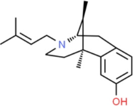
|
• Sigma1 (Kd): 3.9–23.3 nM (Hellewell et al., 1994; John et al., 1999; Colabufo et al., 2004; Azzariti et al., 2006) • Sigma2 (Ki): 1,542–6,611 nM (Hellewell et al., 1994; Vilner and Bowen, 2000; Choi et al., 2001; Ishiwata et al., 2006) |
Agonist (Sigma1) | MTT, MTS, apoptosis assays, light microscopy of cell morphology changes | In most functional studies, it did not impact cell viability or proliferation, and it has been used to block the anticancer actions (cytotoxicity and/or proliferation arrest) of Sigma1 inhibitors/antagonists such as IPAG and rimcazole. In some cases, (+)-pentazocine reported to result in cell detachment and rounding of cells and inhibition of cell proliferation. (3H)(+)-pentazocine is a commonly used radioligand used to quantify and define Sigma1-binding sites. | (Brent and Pang, 1995; Colabufo et al., 2004; Spruce et al., 2004; Rybczynska et al., 2008; Korpis et al., 2014) |
(+)-SKF10047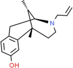
|
• Sigma1 (Ki): 54–597 nM (Hellewell et al., 1994; Vilner et al., 1995a; Ryan-Moro et al., 1996; Vilner and Bowen, 2000) • Sigma2 Ki: 11,170–39,740 nM (Hellewell et al., 1994; Vilner and Bowen, 2000) |
Agonist (Sigma1) | MTT, MTS, or apoptosis assays, light microscopy of cell morphology changes | (+)-SKF10047 has been used to block the anticancer actions (cytotoxicity and/or proliferation arrest) of Sigma1 inhibitors/antagonists such as IPAG and rimcazole. Demonstrated immune modulatory effects by altering cytokine production as well as cytokine-induced signaling in tumor cells. In some cases, (+)-SKF10047 has been reported to result in cell detachment, rounding of cells, and inhibition of proliferation. | (Brent and Pang, 1995; Zhu et al., 2003; Spruce et al., 2004; Do et al., 2013) |
BD1047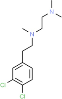
|
• Sigma1 Ki: 0.6–5.3 nM (Matsumoto et al., 1995; Vilner et al., 1995a; Vilner et al., 1995b; Cobos et al., 2005; Entrena et al., 2009) • Sigma2 Ki: 47 nM (Matsumoto et al., 1995) |
Antagonist (Sigma1) | MTS, apoptosis assays, light microscopy of cell morphology changes, in vivo tumor model | Minimal anticancer activity, despite putative antagonist status (defined in behavioral assays). Induced altered cell morphology, but did not cause cancer death. Blocked antiproliferative and cytotoxic actions of Sigma2/TMEM97 ligands. Blocked PRE-084-induced tumor growth in immune competent mouse tumor implantation model. | (Vilner et al., 1995a; Moody et al., 2000; Zhu et al., 2003; Spruce et al., 2004; Kim and Maher, 2017) |
CB-184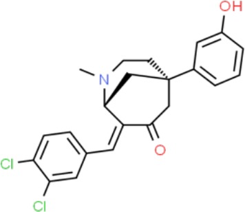
|
• Sigma1 Ki: 7,436 nM (Bowen et al., 1995) • Sigma2 Ki: 13 nM (Bowen et al., 1995) |
Agonist (Sigma2/TMEM97) | MTT, LDH release, apoptosis assays | Cytotoxic effect in cancer cell line cultures as single agent. Potentiated cytotoxic chemotherapeutic agents actinomycin D and doxorubicin. Reported to trigger p53- and caspase- independent apoptosis. | (Bowen et al., 1995; Crawford and Bowen, 2002; Crawford et al., 2002) |
DTG
|
• Sigma1 Ki: 45–203 nM (Hellewell et al., 1994; Vilner et al., 1995a; Vilner and Bowen, 2000; Marrazzo et al., 2011b; Zampieri et al., 2016) • Sigma2 (Ki): 13–58 nM (Hellewell et al., 1994; Vilner and Bowen, 2000; Marrazzo et al., 2011b; Zampieri et al., 2016) |
Agonist (Sigma1 and Sigma2/TMEM97) | MTT, LDH release, apoptosis assays | Blocked voltage-activated K+ currents and induced p27kip1 levels, inhibition of cell proliferation in some studies by proposed G1 cell cycle arrest. Blocked haloperidol-induced cytotoxicity. | (Brent and Pang, 1995; Moody et al., 2000; Colabufo et al., 2004; Renaudo et al., 2004; Kim and Maher, 2017) |
Haloperidol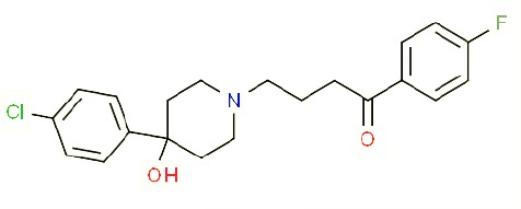
|
• Sigma1 (Ki): 1–40 nM (Vilner and Bowen, 1993; Hellewell et al., 1994; Vilner et al., 1995a; Vilner and Bowen, 2000; Choi et al., 2001; Holl et al., 2009a; Holl et al., 2009b; Holl et al., 2009c; Marrazzo et al., 2011a; Marrazzo et al., 2011b; Weber et al., 2014) • Sigma2 (Ki):12–221 nM Hellewell et al., 1994; Vilner and Bowen, 2000; Choi et al., 2001; Holl et al., 2009a; Holl et al., 2009b; Holl et al., 2009c; Marrazzo et al., 2011a; Marrazzo et al., 2011b; Weber et al., 2014 |
Antagonist (Sigma1) | MTT, MTS, trypan blue exclusion, apoptosis assays, micrographs of cell morphology changes, colony formation, soft agar assay | Antiproliferative and proapoptotic actions in range of cancer cell lines. Reported to induce unfolded protein response and autophagy. Anticancer actions of haloperidol have been proposed to be both Sigma1- and Sigma2-mediated. | (Brent and Pang, 1995; Vilner et al., 1995a; Moody et al., 2000; Colabufo et al., 2004; Spruce et al., 2004; Wang et al., 2004; Nordenberg et al., 2005; Rybczynska et al., 2008; Megalizzi et al., 2009; Sunnam et al., 2010; Pal et al., 2011; Kim et al., 2012; Schrock et al., 2013; Korpis et al., 2014; Kim and Maher, 2017) |
Igmesine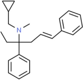
|
• Sigma1 (IC50): 39 nM (Roman et al., 1990) | Agonist (Sigma1) | Trypan blue exclusion, apoptosis assays, cell cycle assays | Inhibited cell proliferation of some cell lines. Blocked voltage-activated K+ currents and induced p27kip1 levels, suggesting G1 arrest. Was not cytotoxic and did not induce caspase-mediated apoptosis. | (Renaudo et al., 2004; Renaudo et al., 2007; Gueguinou et al., 2017; Kim and Maher, 2017) |
IPAG
|
• Sigma1 (Kd): 3 nM (Wilson et al., 1991; Schrock et al., 2013) • Sigma1 low-affinity site (Ki): 500–8,000 nM (Brimson et al., 2011) |
Antagonist (Sigma1) | Trypan blue exclusion, MTT, MTS, CellTiter-Glo, apoptosis assays, cell cycle, soft agar, colony formation assays, in vivo imaging | Selective and potent anticancer activities in range of cancer cell lines, with reported antiproliferative and proapoptotic actions. Induces unfolded protein response and autophagy. Mimics RNAi-mediated knockdown of Sigma1. Triggers lysosomal and proteasomal degradation of cancer promoting signaling proteins including PD-L1, ErbB receptors, and androgen receptor. Multiple high and low-affinity Sigma1-binding sites with distinct activities in intact cancer cells identified. Radiolabeled IPAG tracer used as selective in vivo tumor imaging agent. | (Spruce et al., 2004; Megalizzi et al., 2009; Brimson et al., 2011; Kim et al., 2012; Schrock et al., 2013; Kim and Maher, 2017; Thomas et al., 2017; Maher et al., 2018; Gangangari et al., 2019) |
PB28
|
• Sigma1 (Ki): 10 nM (Azzariti et al., 2006) • Sigma2 (Ki): 0.28 nM (Azzariti et al., 2006) |
Agonist (Sigma2/TMEM97) | MTT, CellTiter-Glo, apoptosis assays, in vivo tumor xenografts | Cytotoxic agent that induces ceramide-dependent/caspase-independent apoptosis in part by triggering the production of mitochondrial superoxide radicals. PB28 also reduced P-gp expression on cancer cell lines. Potentiates doxorubicin. Inhibited tumor growth in vivo. | (Colabufo et al., 2004; Azzariti et al., 2006; Hornick et al., 2010; Hornick et al., 2012a; Hornick et al., 2012b; Niso et al., 2013a; Korpis et al., 2014; Pati et al., 2017a; Kim and Maher, 2017) |
PRE-084
|
• Sigma1 (Ki): 53 nM (Garces-Ramirez et al., 2011) • Sigma2 (Ki): 32,100 nM (Garces-Ramirez et al., 2011) |
Agonist (Sigma1) | Trypan blue exclusion, flow cytometry, tumor allografts | Promoted tumor growth in immune competent mouse tumor allograft model by an IL-10-dependent mechanism. No clear evidence of effects on cancer cell proliferation in cell autonomous culture in vitro or in xenografts. | (Zhu et al., 2003; Kim et al., 2012; Kim and Maher, 2017) |
Rimcazole
|
• Sigma1 (Ki): 406–1,165 nM (Tanaka et al., 1995; Vilner et al., 1995a) • Sigma2 (Ki): 571–852 nM (Schepmann et al., 2011) |
Antagonist (Sigma1) | Trypan blue exclusion, MTT, MTS, CellTiter-Glo, apoptosis assays, cell cycle assays, soft agar colony formation assays, in vivo tumor xenografts | Decreased viability, inhibition of cell proliferation, induction of apoptosis. Inhibition of colony formation in 2D colony formation and 3D soft agar assays. HIF1α induction by rimcazole contributes to its anticancer effects. Inhibited tumor growth and cancer cell proliferation in xenograft studies. |
(Brent and Pang, 1995; Spruce et al., 2004; Achison et al., 2007; Rybczynska et al., 2008; Rybczynska et al., 2013; Happy et al., 2015; Kim and Maher, 2017) |
SA4503
|
• Sigma1 (Ki): 4.6 nM (Lever et al., 2006) • Sigma2 (Ki): 63.1 nM (Lever et al., 2006) |
Agonist (Sigma1) | Trypan blue exclusion, confocal microscopy, in vivo tumor imaging | Blocks IPAG-induced autophagic degradation of PD-L1 in cancer cells. Promotes PD-L1 cell surface expression on cancer cells. (11C)SA4503 development as a tumor imaging agent. | (Ramakrishnan et al., 2013; Kim and Maher, 2017; Maher et al., 2018) |
Siramesine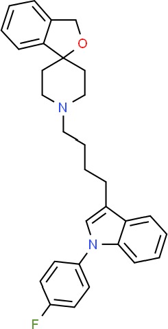
|
• Sigma1 (Ki): 10 nM (Niso et al., 2013a) • Sigma2 (Ki): 13 nM (Niso et al., 2013a) |
Agonist (Sigma2/TMEM97) | MTT, MTS, LDH release, apoptosis assays, in vivo tumor xenograft studies | Lysosomotropic detergent that triggers lysosomal membrane permeabilization and leakage, increased reactive oxygen species, and apoptotic cell death of cancer cells. MEFs transformed with Src or Ras oncogenes sensitized to siramesine-induced cytotoxicity. Inhibited tumor growth in xenograft studies. | (Ostenfeld et al., 2005; Ostenfeld et al., 2008; Hornick et al., 2010; Zeng et al., 2012; Niso et al., 2013b; Zeng et al., 2014; Kim and Maher, 2017) |
SR31747A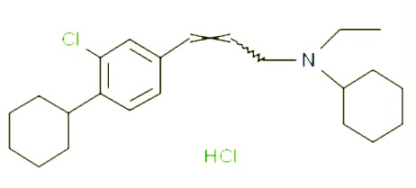
|
• Sigma1 (Ki): 3 nM (Laggner et al., 2005) | Antagonist (Sigma1) | MTT, MTS assays, in vivo tumor xenografts | Immune modulatory and antiproliferative activities. Inhibited proliferation of range of cancer cell lines. Potentiated tumor growth inhibition of flutamide and tamoxifen in xenograft studies. | (Berthois et al., 2003; Ferrini et al., 2003; Casellas et al., 2004; Kim and Maher, 2017) |
SV119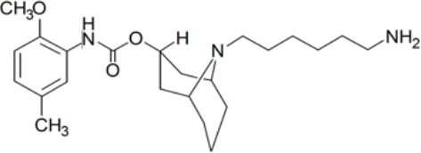
|
• Sigma1 (Ki): 1,418 nM (Vangveravong et al., 2006) • Sigma2 (Ki): 5–8 nM (Vangveravong et al., 2006; Hornick et al., 2010) |
Agonist (Sigma2/TMEM97) | MTS, CellTiter-Glo, LDH release, cell cycle assays, apoptosis assays, colony formation, in vivo tumor xenografts | Inhibited cancer cell proliferation in vitro. Less potent than siramesine. Induced autophagy. SV119 alone induced apoptosis and potentiated cytotoxic and antitumor effects of gemcitabine and paclitaxel in vitro and in xenografted tumors in vivo. | (Kashiwagi et al., 2007; Kashiwagi et al., 2009; Hornick et al., 2010; Spitzer et al., 2012; Zeng et al., 2012; McDonald et al., 2017) |
WC-26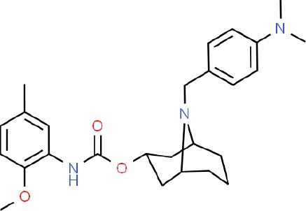
|
• Sigma1 (Ki): 1,436 nM 138 • Sigma2 (Ki): 2.58 nM 138 |
Agonist (Sigma2/TMEM97) | MTS, MTT, LDH release assay, apoptosis assays, colony formation assay | Inhibited cancer cell proliferation and triggered apoptosis in vitro. Induced autophagy. Potentiated doxorubicin-induced cytotoxicity. | (Kashiwagi et al., 2007; Chu et al., 2009; Zeng et al., 2012; McDonald et al., 2017) |