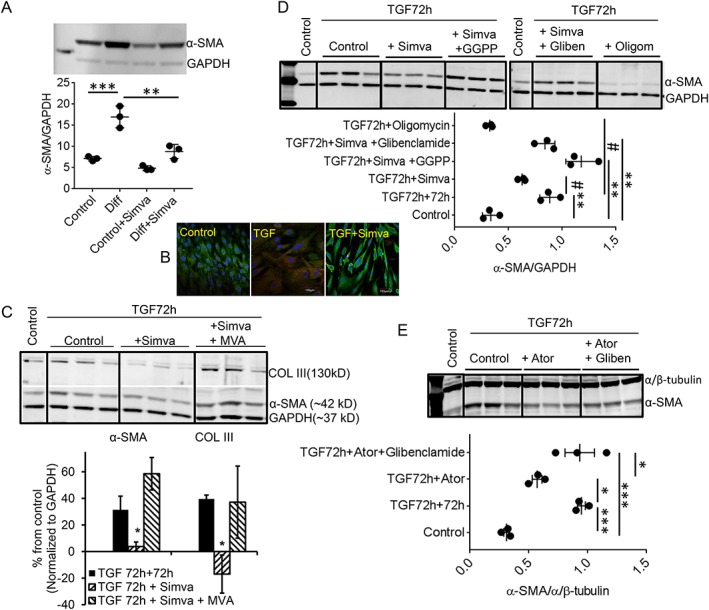
Figure 2.

Mechanisms of statin‐induced de‐differentiation. (A) Representative immunoblot showing the effect of a single concentration of in vitro statin (simvastatin 200 nmol/L for 72 h) on already‐differentiated myofibroblasts (Diff) isolated from HF patients, with significant decrease (64%) in α‐SMA expression (Diff + Simva). (B) Representative immunocytochemistry showed a higher population of α‐SMA+ (red) cells in the differentiated (TGF) cells compared with control or in vitro treatment of simvastatin (TGF + Simva) (green: vimentin). (C) Representative immunoblots from in vitro differentiation. Simvastatin, applied for 72 h following TGF‐β1‐induced differentiation (5 ng/mL for initial 72 h) in vitro (simulating in vivo differentiated hVFs from HF patients), reproduced similar de‐differentiation, as expression of both α‐SMA and COL III were significantly decreased by 87% and 142%, respectively. Mevalonic acid (MVA, 300 μmol/L) prevented the statin‐induced de‐differentiation, as expression of both α‐SMA (31.4 ± 10% vs. 58.6 ± 12%) and COL III (40 ± 3% vs. 37 ± 27%) did not decrease when simvastatin was co‐administered with MVA. (D) Representative immunoblots depict complete prevention of simvastatin‐induced decreased α‐SMA expression by either GGPP (20 μmol/L) or glibenclamide (10 μmol/L). The effect of statin on the expression of a‐SMA is mimicked by oligomycin (1 ng/mL) in the same time frame. Individual data points column graph displays respective mean ratio of α‐SMA to GAPDH densities. (E) Representative immunoblot depicts significant reduction of atorvastatin‐induced decreased α‐SMA expression by glibenclamide (10 μmol/L). Individual data points column graph displays respective mean ratio of α‐SMA to α/β‐tubulin densities. * P < 0.05 vs. control; *** P < 0.001 vs. control; # P < 0.05 vs. TGF‐72 h + 72 h; * P < 0.05 vs. TGF‐72 h + Ator; considered as significant, n = 3 each; one‐way ANOVA. TGF‐72 h + 72 h = TGF‐β1 (5 ng/mL) treatment for 72 h to differentiate into myofibroblasts, and subsequent 72 h culture was without TGF‐β1. Data are mean ± SEM. Ator, atorvastatin; GGPP, geranylgeranyl pyrophosphate; Gliben, glibenclamide; Oligom, oligomycin; Simva, simvastatin.
