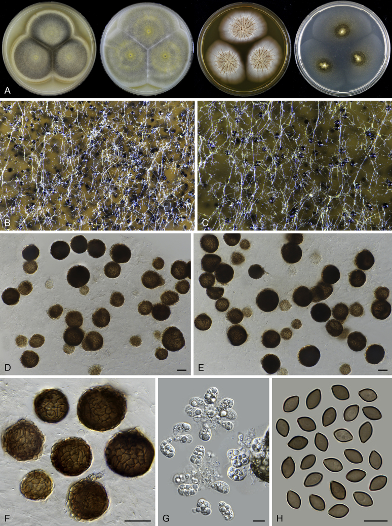
Fig. 32.
Microthielavia ovispora (CBS 165.75, ex-type culture). A. Colonies from left to right on OA, CMA, MEA and PCA after 2 wk incubation. B–C. Part of the colony on OA, showing mature ascomata, top view. D–E. Ascomata mounted in lactic acid. F. Ascomata mounted in lactic acid, showing structure of ascomatal wall in surface view. G. Asci. H. Ascospores. Scale bars: D–F = 20 μm; G–H = 10 μm.

