Figure 1.
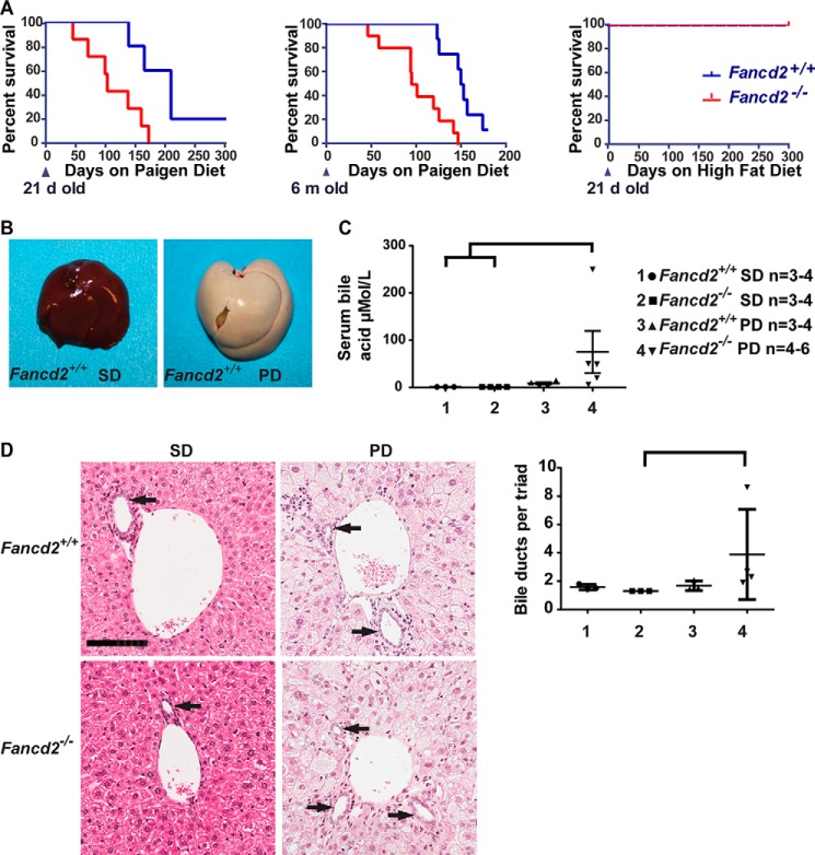
Male Fancd2−/− mice had increased susceptibility to hepatobiliary disease when fed a Paigen diet. A, Kaplan-Meier survival curves showing increased mortality in Fancd2−/− mice fed a Paigen diet beginning at 21 days (left; p = 0.014; Fancd2+/+ = 5 and Fancd2−/− = 8) or 6 months (middle; p = 0.001; Fancd2+/+ = 8 and Fancd2−/− = 10) of age relative to WT mice, and a lack of sensitivity to a high-fat diet (right; p = 1.0; Fancd2+/+ = 8, Fancd2−/− = 5). B, livers were collected from mice fed SD or Paigen diet (PD) for 50–55 days, demonstrating Paigen diet-induced fatty liver disease. There were no grossly detectable differences between genotypes on either diet. C, serum was collected from mice at 50–55 days for blood chemistry analysis using an Abaxis VetScan VS2 chemistry analyzer. Bile acid concentration was significantly increased in male Fancd2−/− mice fed a Paigen diet relative to WT SD–fed mice (p = 0.012) and Fancd2−/− SD-fed mice (p = 0.010). D, hematoxylin and eosin (H&E)-stained liver sections focusing on portal triads. Bile ducts are lined by cuboidal epithelium (arrows). Scale bar equals 100 μm. Quantification of the average number of bile ducts per hepatic portal triad. Fancd2−/− Paigen diet–fed mice had significantly more bile duct profiles than Fancd2−/− SD–fed mice (p = 0.011) indicating Paigen diet feeding induced biliary hyperplasia in Fancd2−/− mice. Error bars represent S.E.

