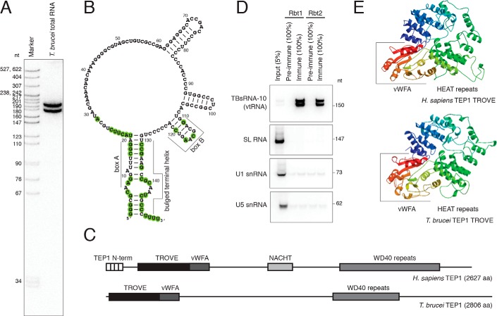
Figure 1.
TBsRNA-10 is the T. brucei vtRNA. A, Northern blot analysis of total T. brucei RNA probed with 32P-labeled antisense TBsRNA-10 oligodeoxyribonucleotide probe. Marker is a 32P-labeled pBR322 DNA MspI digest. B, predicted secondary structure of the T. brucei vtRNA. Shown as double-stranded are only the terminal helix and very stable hairpins emanating from the large internal loop. Evolutionarily conserved residues (see Fig. 3) are highlighted. C, diagram outlining the domain architecture of human and trypanosome TEP1. D, Northern blot analysis of the indicated RNAs after immunoprecipitation with anti-TEP1 sera from two rabbits (Rbt1 and Rbt2). The fractions of the input and precipitated RNA loaded are indicated with percentages. E, three-dimensional structure models for the TROVE domains of human (top) and T. brucei (bottom) TEP1 obtained with SWISS-MODEL (93) based on the crystal structure of X. laevis Ro60 protein (30). The vWFA domains are included in the models.

