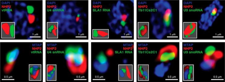
Figure 6.
T. brucei vtRNA is highly enriched in a nuclear compartment distinct from the nucleolus. High-resolution fluorescence in situ hybridization coupled with immunofluorescence was performed for the indicated small RNAs (green) and NHP2 (red), top panels. DNA was stained with DAPI (blue). Bottom panels, simultaneous detection by immunofluorescence of NHP2 (red) and MTAP (blue) with in situ hybridization for the indicated RNAs (green). Insets show three-dimensional representation of the detected RNA and proteins.

