Figure 4.
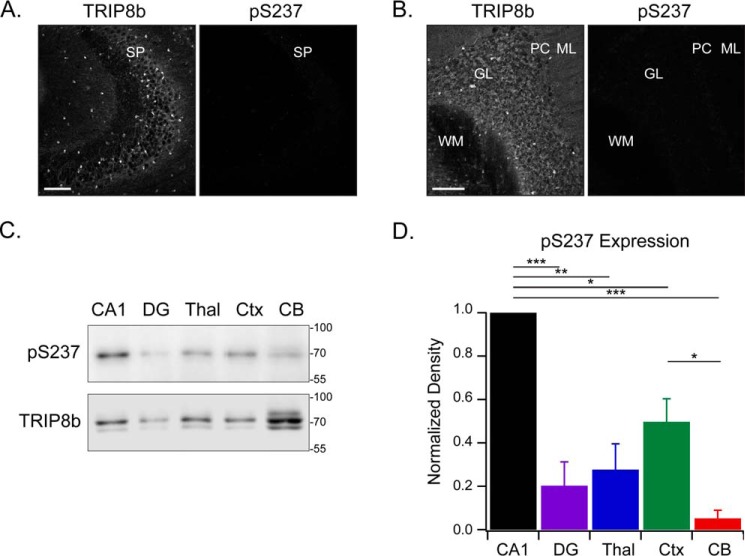
Phosphorylated TRIP8b is absent from the CA3 and cerebellum but highly expressed in the CA1 region of the hippocampus. A and B, CA3 (A) and the cerebellum region (B) stained with antibodies against TRIP8b and pSer237 (n = 3). SP, stratum pyramidale; WM, white matter; GL, granular layer; PC, Purkinje cells; ML, molecular layer. C, protein lysate was loaded onto the gel from various mouse brain regions: 6 μg total lysate from the cornu ammonis 1 (CA1), dentate gyrus (DG), thalamus (Thal), and neocortex (Ctx) and 30 μg of total lysate from the cerebellum (CB). The brain regions were immunoblotted with the indicated antibodies. Note that different quantities of protein had to be loaded to obtain any pSer237 signal, and this led to predictable differences in the total TRIP8b loading control. D, quantification of C, with the pSer237 level normalized to total TRIP8b; one-way analysis of variance with Tukey's post hoc test (CA1, 1.0; dentate gyrus, 0.2 ± 0.1; thalamus, 0.3 ± 0.1; neocortex, 0.5 ± 0.1; cerebellum, 0.05 ± 0.04) with significant differences between the CA1 and dentate gyrus (p < 0.001), CA1 and thalamus (p = 0.001), CA1 and neocortex (p = 0.02), CA1 and cerebellum (p < 0.001), and neocortex and cerebellum (p = 0.03). n = 3; *, p < 0.05; **, p < 0.01; ***, p < 0.001. Scale bars = 100 μm. Molecular mass markers are shown in kilodaltons. All error bars represent mean ± S.E.

