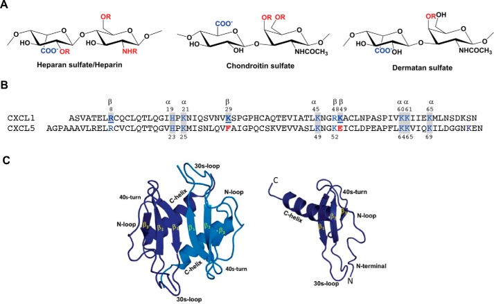
Figure 1.
A, schematic of heparin/HS, CS, and DS structures. R stands for potential sulfation sites. B, sequences of CXCL1 and CXCL5. Basic residues implicated in GAG binding that are also conserved in other neutrophil-activating chemokine are highlighted in blue and shaded in gray, those that are involved in GAG binding in CXCL1 alone are in bold and underlined, and corresponding residues that are not conserved in CXCL5 are in red. C, structures of CXCL1 dimer and monomer. All CXCR2-activating chemokines share high sequence similarity and have the same structural fold. The individual monomers in the dimer are shown in dark and light blue for clarity. Different structural and functional regions are labeled in both the monomer and dimer.

