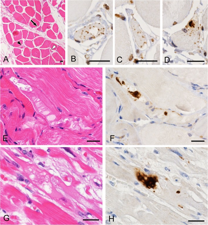
Fig. 6.
Contiguous sections of the cervical muscle (a-d), diaphragm (e and f) and myocardium (g and h) stained with HE (a, e and g) and anti-pTDP-43 (b-d, f and h). a-d Muscle fibers showing single-fiber atrophy (a) contain pTDP-43-positive short linear inclusions (b and c: black and white arrowheads in (a)) and dense filamentous inclusions (d: arrow in (a)) in a patient with non-neuromuscular disease (case B-47). Note that atrophic fibers contain small vacuoles (b). e and f A muscle fiber showing marked vacuolar degeneration (e) contains dense filamentous inclusions immunopositive for pTDP-43 (f) in a patient with ALS (case B-16). g and h A cardiac muscle fiber showing marked vacuolar degeneration (g) contains dense filamentous inclusions immunopositive for pTDP-43 (h) in a patient with Duchenne muscular dystrophy (case B-37). Bars = 20 μm

