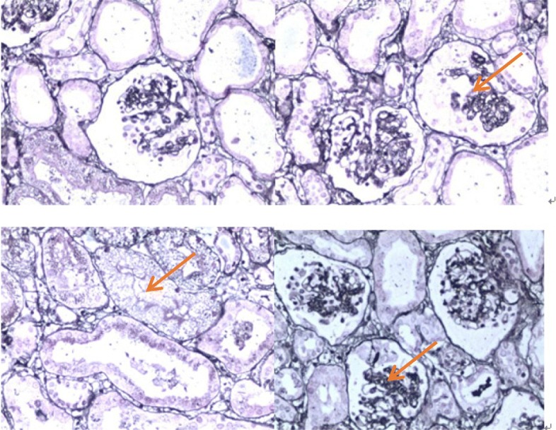
Fig. 1.

Renal histology and ultrastructural findings in light microscopy. It showed 48 glomerular, 4/48 were immature glomerular; 17/48 had glomerular mesangial cells and matrix hyperplasia with different levels of insert appearance (glomerular collapse, did not see open capillary lumens); 15/48 had glomerular balloon expansion, the expansion of multiple focal proximal convoluted tubules, renal tubular epithelial lesions sliced and interstitial oedema; and small arteries did not show obvious pathological changes. Immunohistochemical tests results of 4 glomerular sections: IgG(−), IgA(−), IgM(−), C3(−), C1q(−), and Fn(−) (haematoxylin and eosin stain). Magnifications: × 200
