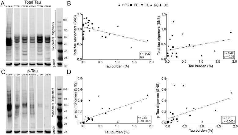
Fig. 3.
Representative images of Western Blot membranes probed with total tau (a) and PHF-1 (c) antibodies showing lower amounts of p-tau monomers and oligomers in CTE compared to AD brain samples. Significant correlations were found between total tau burden quantified on immunostained sections and levels of tau oligomers (b) and p-tau monomers and oligomers in CTE cases (d). Spearman r and p values are displayed on the graphs. Abbreviations: HPC: hipoccampus; FC: frontal cortex; TC: temporal cortex; PC: parietal cortex; OC: occipital cortex

